I used Gemini Notebook to generate a 50-minute audio overview of this file: https://
See the clot3.
The sections of this file are sorted in ascending chronological order, and they have been added between February and October of 2026.
Kevin McCairn has developed a lucrative business where he tricks people into thinking that they have amyloid fibrin microclots in their blood, and then he charges people 60,000 USD to remove the clots.
The structures he typically claims are fibrin microclots are string-like fibers with a diameter around 10-30 µm. In reality they are likely to be fibers from dust, like textile fibers or paper fibers. Cellulose fibers have a diameter around 10-40 µm, including cotton fibers and paper fibers. Nylon fibers similarly have a diameter around 10-50 µm, and polyester fibers have a diameter around 10-15 µm.
Previously fibers in blood samples with a diameter around 10-30 µm have been called graphene oxide ribbons by Philippe van Welbergen, hydrogel filaments by Ana Mihalcea, Morgellons filaments by people in Arne Burkhadt's pathology conference, and fiber optic killer tubes by Shimon Yanowitz.
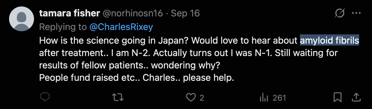
McCairn used to say that the fibers he saw under a microscope were amyloid fibrils, but he dropped the term fibril because I told him that the term fibril refers to nano-scale structures in biology, and amyloid fibrils have a diameter around 7-13 nm, so they are not even visible with an optical microscope.
The flat ribbon-like shape of McCairn's fibers is also consistent with textile fibers but not with fibrin. Some of his fibers have had sharp corners similar to paper folds, where you can see one part of a flat fiber folded over another part, which is consistent with synthetic textile fibers but not with fibrin.
McCairn does not determine the actual protein composition of his fibers using methods like immunostaining, spectroscopy, or proteomics. But he jumps to the conclusion that if a fiber is fluorescent under a UV light, then the fiber is made of amyloid fibrin, even if the fiber was simply autofluorescent without staining by Thioflavin T. But for example white textiles usually contain optical brightening agents which are fluorescent under UV light. The microclot researcher Resia Pretorius pointed out that white tissue paper fibers are similarly fluorescent, and she indicated that some of the fibers McCairn identified as amyloid clots looked like false positives.
McCairn tells people that when they mail him a sample of blood, they should spread out a drop of blood on a glass slide and let the blood dry overnight in a dust-free environment. But how many people have a perfectly dust-free environment at their home? He specifically tells people to not place a cover slip over the blood, so that the blood remains exposed to open air. And he doesn't tell people to check the next morning that no dust fibers fell on the blood while it was drying.
A fiber with a diameter of 15 or 30 µm couldn't even be circulating freely in human blood, because the fiber would be too big to pass through the capillaries. Capillaries typically have a diameter around 8 µm, which is barely wide enough for a red blood cell to pass through, but the fibers shown in McCairn's microscope images have often been about 2 to 4 times wider than red blood cells.
Even if the fibers with a diameter of 10-30 µm were circulating in blood, the fibers would not be removed by the plasmapheresis ciruit used at Edogawa Hospital. The first filter in the circuit is a plasma separator, which separates the plasma from the red blood cells. The separator has a pore size of 0.3 µm, which is too small for red blood cells to pass through, so the red blood cells get returned to the patient's blood without any filtering. Only components of blood that are small enough to pass through the pores get routed to the immunoadsorption column, which is the filter in the circuit that is meant to provide the therapeutic benefit. However fibers with a diameter of 10-30 µm would be far too big to pass through the pores of the plasma separator, so the fibers would simply be returned back to the patient's blood without any filtering, like what happens to the red blood cells.
The treatment provided by Edogawa Hospital is often incorrectly called double filtration plasmapheresis (DFPP), but in reality the treatment is immunoadsorption (IA), which is also known as plasma adsorption (PA) in Japan. DFPA appears to be a nonstandard term for IA invented by Edogawa. McCairn initially referred to the treatment as DFPP, but later he switched to calling the treatment DFPA, which he says means "dual filtration plasma apheresis". So confusingly the acronym DFPA has nearly the meaning as DFPP, but with "plasma apheresis" written as two words, and not in the more common form of "plasmapheresis" as a single word. In Japanese-language materials, Egogawa refers to the treatment as PA, or plasma adsorption, and sometimes the letter A in DFPA is also said to stand for "adsorption" and not "apheresis".
The nonstandard term DFPA makes the treatment provided by Edogawa appear somehow unique, because treatment called DFPA is not available outside of Edogawa, even though in reality DFPA refers to regular immunoadsorption treatment that is widely available outside of Japan. The TR-350 column used by Edogawa is distributed in Europe by a German company called Diamed.
In a German RCT of immunoadsorption as a treatment for long COVID, no significant benefit was found compared to a sham treatment, even though the trial employed the same Asahi TR-350 immunoadsorption column that is used by Edogawa Hospital.
McCairn used to say that the fibers he saw in blood samples under a microscope were "amyloid fibrils", but after I told him that amyloid fibrils are nano-scale structures, and the term "fibril" in general refers to fibers with a nano-scale diameter in biology, he started saying that the fibers were "fibrillar formations" instead (which was still somewhat confusing, because some people continued to interpret the term as meaning that the fibers were amyloid fibrils). Now he seems to have invented another new term for the fibers, and he has started to call them "aggregates".
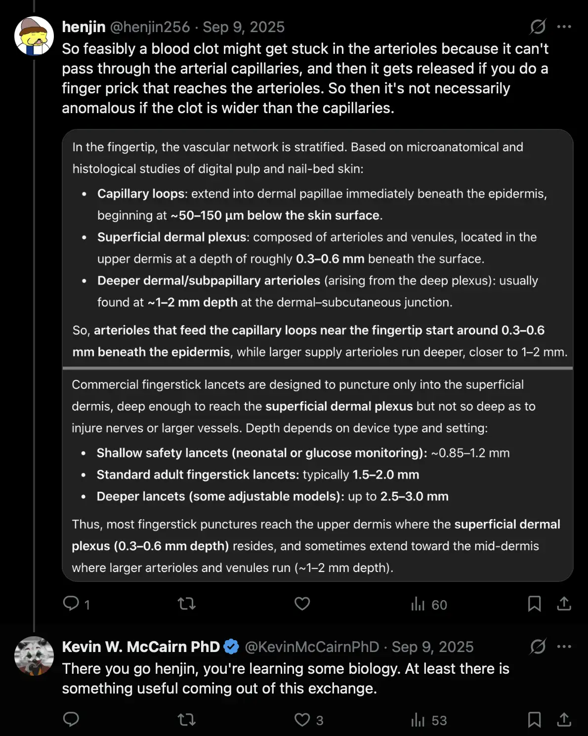
Earlier McCairn gave the impression that the fibers he found in blood samples were circulating freely in the bloodstream, but me and another user on his Discord pointed out that his fibers had a wider diameter than capillaries, so the fibers wouldn't be able to pass through the capillary bed. But then later I figured out that in case the fibers were lodged in the arterioles before the capillaries in the fingertips, the fibers might get released from the arterioles if you do a finger prick sample, because typically a finger prick lancet would reach deep enough to penetrate the arterioles and not only the arterial capillaries. However it's a hypothesis I came up with on my own and that McCairn didn't suggest to me (and I came up with the hypothesis because I was trying to play along with the scenario where the fibers that look like textile fibers would actually be clots made of fibrin, which I was nearly certain was not true): [https://
Earlier I asked McCairn several times if the reason why his supposed clots had a string-like shape was because the clots formed in a small blood vessel and then got dislodged, McCairn came up with explanations like that "there is a coherency in the epistemological grounding of the amyloid PRION formation". [https://
However now when he did a video with one of his first patients, McCairn said himself that the so-called "aggregates" are "lodged into the entrance and exits of capillary beds", where he even used the same verb "lodged" that I had earlier used many times to refer to the hypothetical clots that are stuck before the capillaries. He said: "So, what's the one thing that we can point at right now, which is quantitative in a ways that the aggregations in the plasma disappear? And, are they generated de-novo, or are they a consequence of initial exposure, and then just sort of building up over time? I'm still unsure. But what I can say is between the DFPP - PA session, sorry - you were in the second treatment protocol, in the pre-plasma - there was amyloid-like burden in the pre-plasma that has gone in the post. So my thinking is that there's a bulk movement of the plasma during filtration, and that's gonna drag a whole bunch of these aggregates with them. It's coming back in clean. And then if there's more that are sort of hanging on to the vasculature, they're gonna then be washed off into the plasma again, which is why we're seeing the return of them in the secondary treatment - sorry, in the second treatment. And I would say the fact that you've such a rejuvenating effect - those are the aggregates that were really lodged into the entrance and exits of capillary beds. And so, I think we're moving them, but like I'm saying, in your case it's taking longer than two weeks." [https://
But if the aggregates are lodged before the capillaries but the aggregates are too big to pass through the capillaries, then how could the aggregates be removed by the plasmapheresis treatment? And if the aggregates are lodged after the capillaries, then how do they happen to break loose at the same time when the patient undergoes the plasmapheresis treatment? Does the treatment include some mechanism that induces the aggregates to become embolized?
As of April 2026, McCairn's hardcore fan CoyoteSanctuary was still using the term "amyloid fibril" to refer to the structures McCairn sees in blood samples (or actually he used the word "fibrilla", but "fibrilla" is synonymous with "fibril"): [https://

Added in September 2026: One of McCairn's early patients still uses the term "amyloid fibril" to refer to the fibers McCairn finds under a microscope: [https://
McCairn supposedly found this fiber in the blood of one his patient Ken Evans, whose username on Twitter is KenCaptn20114: [https://
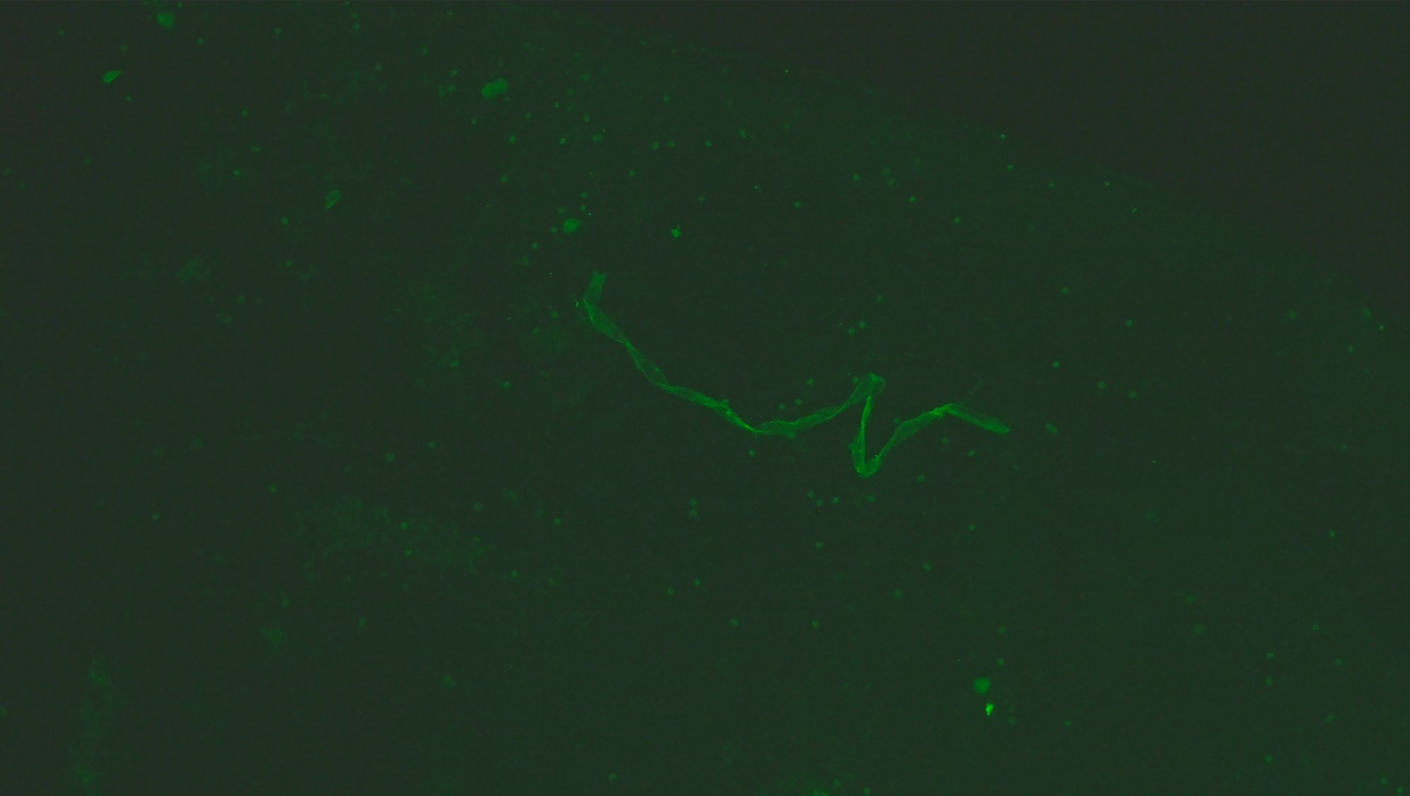
If the fiber is a fibrin clot that has a string-like shape because it formed inside a blood vessel so it conformed to the shape of the blood vessel, then why is the fiber flat and not cylindrical?
This is what ChatGPT said about the fiber:
What you're seeing is almost certainly a contaminant fiber, not a biological structure from the blood itself.
A few cues from the image:
Uniform thickness along most of its length → biological filaments (like fibrin strands) tend to branch irregularly and form mesh-like networks, not long smooth ribbons.
Sharp bends/kinks → typical of synthetic or textile fibers that have been mechanically deformed.
Flat, ribbon-like morphology → very characteristic of textile fibers (e.g., cotton, rayon, or synthetic microfibers), which often appear twisted or collapsed into ribbon shapes under microscopy.
Strong fluorescence under UV → many fibers (especially synthetic ones or those treated with dyes/brighteners) fluoresce brightly, whereas native blood components usually don't show this kind of linear, high-contrast emission unless specifically stained.
Most likely candidates
Cotton fiber (common lab contaminant; appears as a flattened, twisted ribbon)
Synthetic microfiber (polyester/nylon; often smoother and more uniformly fluorescent)
Lint contamination from clothing, wipes, or lab materials
What it is not likely to be
Fibrin strand → would look more web-like and irregular
Parasite or worm → would show internal structure and biological segmentation
Cellular debris → would be more granular or amorphous
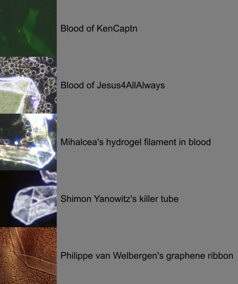
The fiber has one of these "paper fold"-style corners where you can see folded over segments of the flat fiber overlapping each other, like in the case of the fiber in the blood of Jesus4AllAlways, Mihalcea's hydrogel filament, Shimon Yanowitz's killer tube, and Philippe van Welbergen's graphene ribbon:
KenCaptn said that the treatment he got in Japan was very expensive, but he had no choice because he would've died if he didn't get the treatment: [https://

He tweeted: "This clot issue possibly could turn into the biggest problem that humanity has ever faced. Think youre safe because youre unvaxxed? Think again. There are violently ill people here that are unvaxxed in Japan covered in microclots being treated." [https://
KenCaptn also said that stem cell growth factors repair demons: [https://

He said that blood clots are "called fibril or amyloids": [https://

He claims that his life was earlier saved by Mary Talley Bowden who suggested him to double his dose of ivermectin and nattokinase: [https://
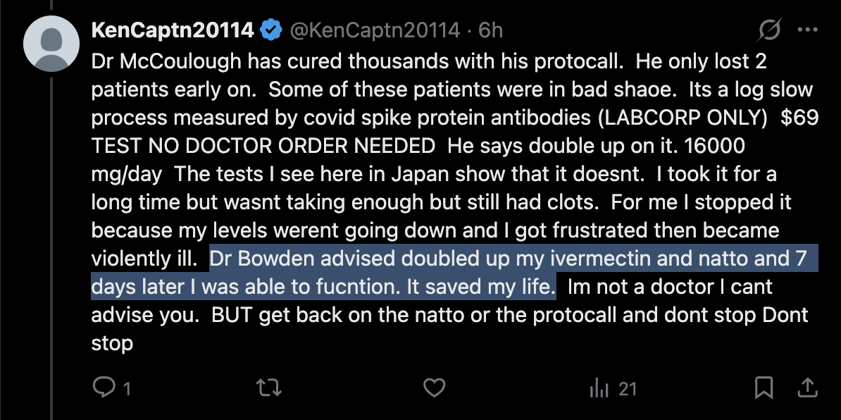

Even after he had received the plasmapheresis treatment, he still said that he is afraid to ever get off ivermectin: [https://

Now McCairn seems to have come up with a new explanation for why some of his supposed clots have a flat and not cylindrical cross-section, which is that "veins are flat and non-cylindrical": [https://
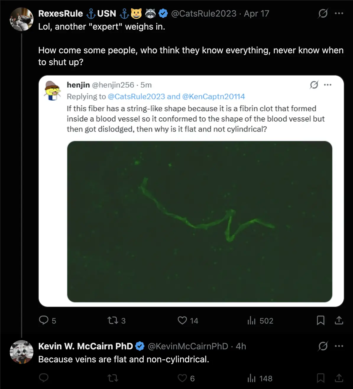
So he seems to still be saying that the shape of the clot is explained by the clot being an intravascular cast. ChatGPT said:
Short answer: generally no - a fibrin clot that detaches from a vessel does not reliably preserve the vessel's cross-sectional shape, especially not as a stable, flat "ribbon."
Fibrin networks are viscoelastic and deformable rather than rigid. Once a clot is dislodged and no longer constrained by blood flow and vessel walls, it undergoes stress relaxation, elastic recoil, and internal restructuring. As a result, it can collapse, twist, or fragment, rather than maintaining a fixed molded geometry.
Although veins can be partially collapsed or non-circular in vivo, particularly at low pressure, their lumen remains a three-dimensional, compliant structure rather than a consistently flat channel. After removal from the body, the loss of internal pressure and surrounding tissue support leads to further deformation. Additional handling and fluid forces during extraction can significantly alter the clot's shape.
There are cases where biological material approximates a lumen, such as airway casts or some post-mortem clots, but these typically appear as irregular cylindrical or branching structures. They do not present as long, uniformly flat ribbons.
A persistent flat, ribbon-like morphology is therefore more consistent with ex vivo artifact, mechanical compression, surface-interface effects, or non-fibrin material. It is not well explained by simple intravascular molding of a fibrin clot.
Overall, while a clot may transiently resemble the vessel it formed in, it does not stably retain that geometry, and a consistently flat cross-section is not what would be expected from a true intravascular fibrin clot.
When I asked RexesRule why KenCaptn's fiber was flat and not cylindrical, she didn't answer my question, but she just said that I was a troll, and that McCairn and his patients are heroes who don't deserve trolls in their threads: [https://

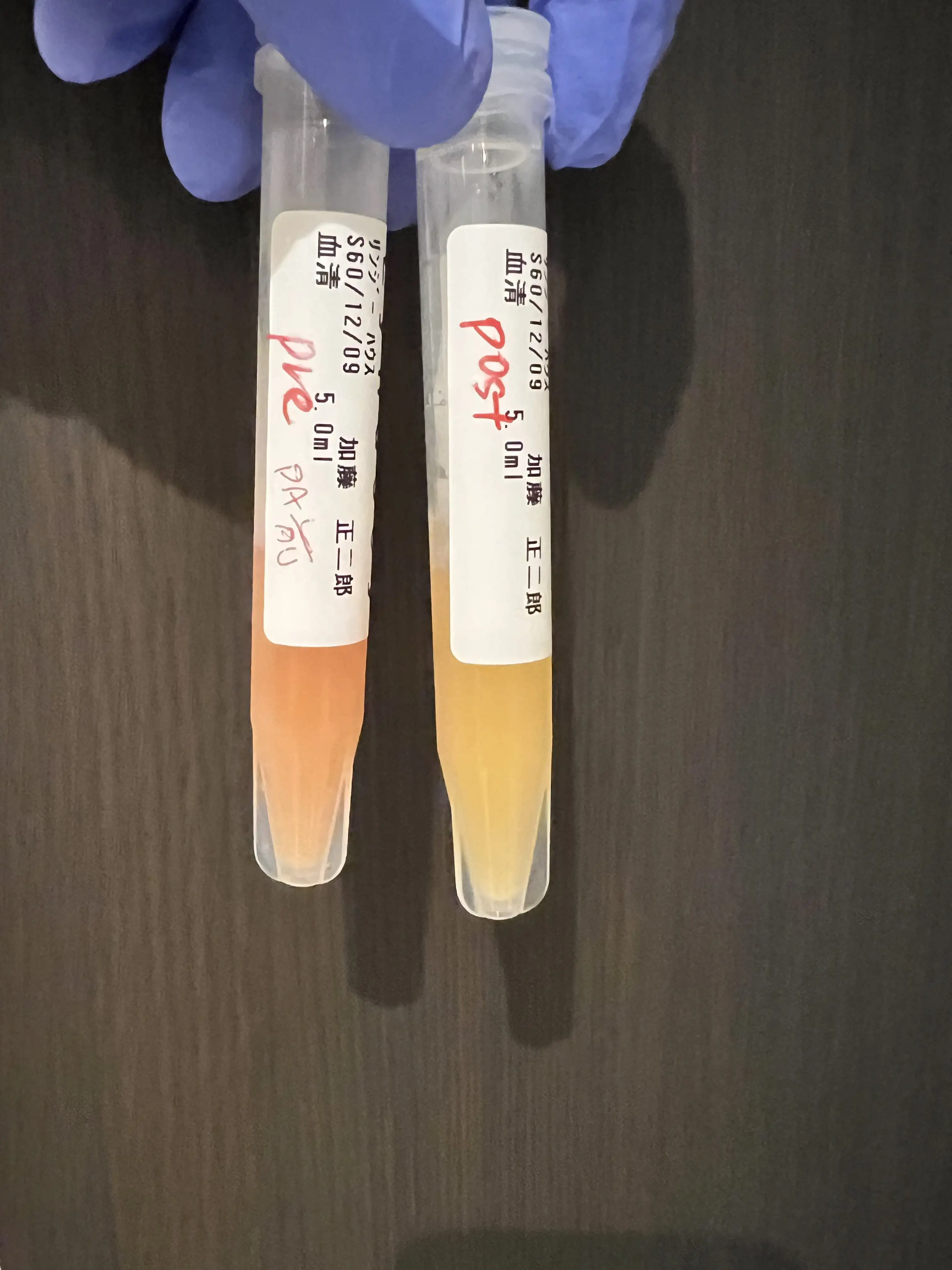
Lyndsey House showed that her plasma was more reddish before she got immunoadsorption treatment and more yellowish after the treatment, as if that was supposed to show that the treatment was somehow effective: [https://
Another patient showed that her plasma was more yellowish before treatment but more reddish after treatment: [https://
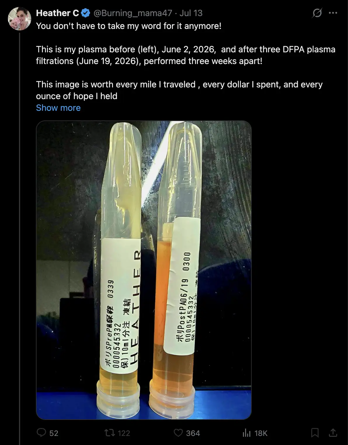
Reddish color in plasma is typically due to hemolysis, where hemoglobin has been released to the blood from ruptured red blood cells. So the change in the color of the plasma is not necessarily due to the effect of the treatment. ChatGPT said:
The color of plasma in photographs is not determined by a single substance, so a change from yellowish to reddish—or the reverse—does not by itself indicate that immunoadsorption removed or added a particular type of material.
Yellow or straw-colored plasma is the normal appearance of relatively clear plasma. Its color is largely due to bilirubin and other naturally occurring pigments, with albumin and other plasma constituents affecting the optical properties. A stronger yellow color can therefore reflect ordinary biological variation, including bilirubin concentration, rather than a pathological component.
Reddish or pinkish plasma usually indicates some degree of hemolysis: red blood cells have ruptured and released hemoglobin into the plasma. Even a modest amount of free hemoglobin can make plasma visibly pink, red, or orange. Importantly, hemolysis can occur during blood collection, centrifugation, transport, or processing, so a reddish post-treatment sample does not necessarily mean that immunoadsorption itself caused the change.
This is one reason the direction of the color change can differ between patients or photographs. If the pretreatment sample happened to have more hemolysis than the post-treatment sample, it could become less reddish after treatment and appear relatively yellow. Conversely, if hemolysis occurred during or after the treatment sample was collected, the post-treatment plasma could become more reddish. Differences in bilirubin, lipids, medication pigments, sample handling, lighting, camera white balance, tube material, and background can also change the apparent color.
Lyndsey and another one of McCairn's patients reported reduced numbness in their toes after getting the DFPP treatment, which might be an outcome of improved perfusion to the toes: [https://

I was wondering that if DFPP doesn't actually induce microclots lodged in the capillaries to break loose, then how could DFPP improve perfusion to the toes? Grok speculated that DFPP might improve perfusion by reducing the viscocity of blood:
Fibrinogen is a key high-molecular-weight plasma protein that dramatically increases viscosity (especially in low-shear microcirculation). DFPP removes 50-88% of fibrinogen per session or cumulatively (along with other contributors like IgM, α2-macroglobulin).
Lower viscosity directly increases capillary flow rate per Poiseuille's law: flow ∝ (pressure × radius⁴) / (viscosity × length). In narrow capillaries, even small viscosity drops yield large perfusion gains. This improves oxygen exchange without needing to dissolve every lodged clot. Studies on plasmapheresis in hyperviscosity syndromes confirm this: reduced plasma viscosity enhances capillary perfusion, shear-dependent vasodilation (via endothelium-derived relaxing factors), and tissue oxygenation.
Result: Better overall microcirculatory flow, even around partial obstructions, leading to the "rejuvenating effect" described in patient reviews.
A paper about HELP apheresis by Beata Jaeger et al. said: "In principle, the decreased fibrinogen concentration causes rheologically significant effects and facilitates oxygen exchange. Plasma viscosity is reduced by an average of 19%, and erythrocyte aggregability is significantly decreased by 60% (33). In addition, the vascular endothelial growth factor (VEGF) and nitric oxide (NO) release are favorably influenced (33). The improvements have also been demonstrated for cerebral blood flow in the cardiac patients, where they profit from a 63% increase in the CO2 reserve capacity (54)." [https://
The reduction in peripheral neuropathy might also be explained by a neurological and not circulatory mechanism. Plasmapheresis is employed as a treatment for chronic inflammatory demyelinating polyneuropathy (CIDP), which is an autoimmune condition that affects the myelin sheath around peripheral nerves. Plasmapheresis removes antibodies in plasma that attack the nerves, even though the effect of the treatment typically lasts for only a few weeks, so the treatment has to be repeated frequently. [https://
A paper from 2011 said: "Based on 2 Class I studies, plasmapheresis is established as effective in the short-term treatment of CIDP; both studies showed the beneficial effect is not sustained, with worsening beginning 1-5 weeks after last plasmapheresis treatment." [https://
When I asked what other explanations there might be for the reduced numbness in the toes after the DFPP treatment, Grok produced this list of 4 possible explanations:
1. Reduction in systemic inflammation and neurotoxic/inflammatory mediators (non-CIDP pathway)
DFPP removes cytokines (e.g., TNF-α), chemokines (e.g., RANTES), oxidized LDL, complement components (C3/C4), and other pro-inflammatory molecules that can directly sensitize or damage peripheral nerves or cause small-fiber neuropathy symptoms.
In patients with metabolic or non-metabolic peripheral neuropathy (including cases with environmental toxin burden), DFPP has been shown to significantly lower serum CRP, TNF-α, eosinophilic cationic protein, and fibrinogen while simultaneously removing heavy metals/pesticides - correlating with clinical sensory improvement.
Similar rapid anti-inflammatory effects are reported in autoimmune encephalitis and other neuroinflammatory states treated with DFPP, where neurological function (including sensory symptoms) improves even without classic demyelinating neuropathy.
This is a neurological (rather than purely circulatory) mechanism: the nerves themselves are less irritated or inflamed, independent of blood flow.
2. Improved rheology beyond simple viscosity (erythrocyte behavior, endothelial function, and microcirculatory dynamics)
Even without physically "breaking loose" lodged microclots, DFPP alters blood flow properties in ways that enhance capillary perfusion and tissue oxygenation:
It reduces erythrocyte aggregability/rouleaux formation (by ~60% in analogous HELP apheresis) and lowers plasma viscosity more broadly than fibrinogen removal alone. This increases shear stress on vessel walls, triggering endothelium-derived relaxing factors (nitric oxide, VEGF) that promote vasodilation.
In conditions like arteriosclerosis obliterans (ASO) or diabetic foot syndrome - where patients classically report toe numbness, coldness, and pins-and-needles - DFPP is an established therapy precisely because it improves lower-limb microcirculation and sensory symptoms via these rheological changes.
Long-COVID apheresis studies (using similar filtration) document disappearance of fibrin fibers and rouleaux on dark-field microscopy alongside symptom relief, showing flow improvement without requiring clot dissolution.
Poiseuille's law still applies, but the effect is amplified by better red-cell deformability and reduced cellular aggregation, allowing plasma to "squeeze" through partially compromised capillaries more effectively.
3. Removal of other hyperviscosity or pro-aggregatory factors (e.g., cryoglobulins, lipids, immune complexes)
DFPP is highly effective for cryoglobulinemic vasculitis or hyperviscosity syndromes, where large proteins cause sluggish flow and neuropathy; sensory symptoms (including distal paresthesias) often resolve rapidly after sessions.
Even in non-cryoglobulinemic cases, it removes oxidized lipids and immune complexes that promote endothelial dysfunction or microvascular sludging - effects documented in long-COVID cohorts and peripheral-artery-disease patients.
Grok said that blood viscosity would typically return close to a normal level in 3-6 days, fibrinogen in 2-5 days, cytokines like TNF-α and IL-6 in about 1-2 days, and oxidized LDL in 1-7 days.
(Edited later: The treatment provided at Edogawa is not actually DFPP but immunoadsorption, but the comments above are also to some extent applicable to immunoadsorption.)
The German anti-vaccine researcher NarfGb posted these comments about McCairn's treatment protocol: [https://
Sorry to say, but this sounds like voodoo without any given evidence! Treating symptoms temporarily is not healthy, offers no better survival chances, and is nothing more than voodoo medicine.
More open questions than anything I'd suggest for treating:
How was it proven that apheresis triggered anything more than a temporal and/or placebo effect? (How was a sham-controlled effect size established to differentiate therapeutic apheresis from a temporal/ placebo response?)
How was the clearance of modRNA determined following apheresis? (Lack of pharmacokinetic data regarding the removal of synthetic transcripts from systemic circulation.)
In the event of persistence, how were integrative events and/or RNA turnover, driven by cellular homeostatic effects, excluded? (Addressing the risk of genomic integration or compensatory mechanisms masking the treatment's actual impact.)
How was the clot formation mechanistically attributed to modRNA/Spike persistence without relying on third-rate in vitro experiments that lack any reliable diagnostic or predictive value?
How was the individually dependent dosage determined, particularly concerning the administration of growth factors? (Considering the non-linear dose-response curve and the risk of biological recklessness.)
Which specific exosome formulation was utilized? (Demanding CMC, Chemistry, Manufacturing, and Controls-data: source, purity, and surface markers.)
Which specific growth factors were used, and what specific cell-line was their recombinant or biological origin? In other terms: what is "SHED"?: It is not clear if it were Mesenchymal stem/stromal cells (Dental Pulp MSCs / SHED-MSCs), Perivascular stem cells / pericyte progenitors, Neural crest-derived stem/progenitor cells, Neural stem or neural progenitor cells, Odontoblastic progenitor cells (dentin-forming precursors)
How was the individual dose-dependency established to avoid over-stimulation of signaling pathways?
By what methodology were the exosomes tracked in vivo to confirm biodistribution and target-tissue engagement?
How were off-target effects and non-specific cellular interactions rigorously excluded?
What is the clearly defined molecular mechanism of action (MoA), including validated in vivo dose-response relationships, that substantiates causality beyond descriptive reports and in vitro experiments employing recombinant spike proteins lacking physiologically relevant dosing?
Where is the supporting multi-omics data (transcriptomics, proteomics, metabolomics, lipidomics (due to using exosomes)) to provide a quantifiable biological basis?
How were all other potential etiologies, other than the "Spike protein", differentially excluded?
What is the exact N-number and stratification of the cohort regarding previous modRNA-LNP injections?
How was electrostatic membrane interference and receptor shifting excluded, which may lead to disturbed intra-cellular communication, as primary drivers for thrombotic events?
How is the absence of time-shifted feedback loops or paradoxical immune responses excluded after application that could lead to long-term exacerbation?
Without addressing dose-dependent toxicity and providing longitudinal omics-data, and a clear tier of evidence this protocol remains a speculative 'Black Box' that fails the most basic requirements of the Helsinki Declaration.
In the comment section NarfGb wrote: "There is not even any animal experiments done. But using it for people with real issues and getting paid for doing human experiments you think is a good thing?! Especially with a therapy that does not really differ from LNPs despite a little bit more biocompatible lipid composition?"
He also wrote: "Sadly: It is not even a scientific argumentation: 'BUT they look and feel great.' I feel also great smoking crack or taking steroids." And he expressed concern how McCairn did not provide numeric data to quantify the effect of his treatment, but he presented evidence like an infomercial-style image where the patient looked happier in the "after" picture than the "before" picture. [https://
NarfGb also wrote: "And in the first line I am more pissed about the fact that he sells this protocol as a mirrage cure than to say it's pure experimental and we don't know by now what we're doing. This is the part really freaks me out." I agree with his concern, and furthermore McCairn has been fearmongering about the clots to the extent that some of his followers now believe that the clots are leading to human extinction. One of his patients said that the clots might be the most important issue humanity is facing, and that everyone is dying from the clots, and that he would have died if he didn't get McCairn's treatment. The patient also speculated that the stem cell growth factors "repair demons", which makes it seem like he didn't understand the purpose of the treatment well enough to have informed consent.
NarfGb next published a second post about the McCairn's protocol. [https://
I'm not saying that abnormal, fibrinolysis-resistant clots don't exist. The fluorescence signal is reproducible. But the term "amyloid" is overinterpreted and unproven.
I replied:
I agree, and the "amyloid" terminology fuels McCairn's fearmongering about how the vaccines are causing a prion zombie apocalypse.
Two years ago Baxter Dmitry from The People's Voice published an article that said: "According to Dr. McCairn, contagious prions released by Covid jabs behind the explosion of disease around the world and explain the mechanism for phenomena including 'silent hypoxia', the formation of white fibrous clots, the sudden deaths of athletes, turbo CJD, turbo cancer, amyloidosis, and the explosion of neurological diseases." [https://
thepeoplesvoice. ]tv/ neuroscientist- contagious- prions- from- covid- jabs- are- transfecting- the- unvaccinated/
Then NarfGb told me:
You knew, that you'll find in almost every protein amyloidogenic peptide sequences?
Tzotzos S, Doig AJ. Amyloidogenic sequences in native protein structures. Protein Sci. 2010 Feb;19(2):327-48. doi: 10.1002/pro.314. PMID: 20027621; PMCID: PMC2865711.
That was the best joke for the panic narrative: using the worst sequences to sell a panic narrative and distract from the injections.
I told him that was a great point, and I hadn't explored that angle before. Grok said:
Amyloidogenic or aggregation-prone sequences (APRs) are very common across proteins. These short segments, typically 5-15 residues long and often hydrophobic with high β-sheet propensity, occur frequently in polypeptide chains. Predictive tools such as TANGO, WALTZ, and AGGRESCAN, along with large-scale proteome analyses, indicate that APRs appear on average at least once every ~50-100 amino acids in typical protein sequences. In globular proteins, roughly 15-20% of residues fall within such predicted aggregation-prone regions, though these are usually buried in the hydrophobic core or stabilized by native folding to prevent uncontrolled aggregation. [https://
www. ]nature. com/ articles/ s41467- 020- 17207- 3 Proteome-wide surveys confirm this high prevalence. In a comprehensive analysis of plant proteomes spanning 75 species and more than 2.9 million protein sequences using the WALTZ algorithm, more than half of all proteins in each proteome contained at least one predicted amyloidogenic region, with a median of around 25% of proteins after applying stricter filters. [https://
www. ] Similar patterns hold in other organisms, including bacteria and eukaryotes. Many proteins - perhaps most - harbor the potential to form amyloid-like structures under permissive conditions such as fragmentation, denaturation, high local concentration, or specific environmental stresses.mdpi. com/ 1422- 0067/ 18/ 10/ 2155
A user that promotes Miles Guo said that the CCP is using a protocol similar to the McCairn protocol but with fresh blood taken from PLA military boys: [https://

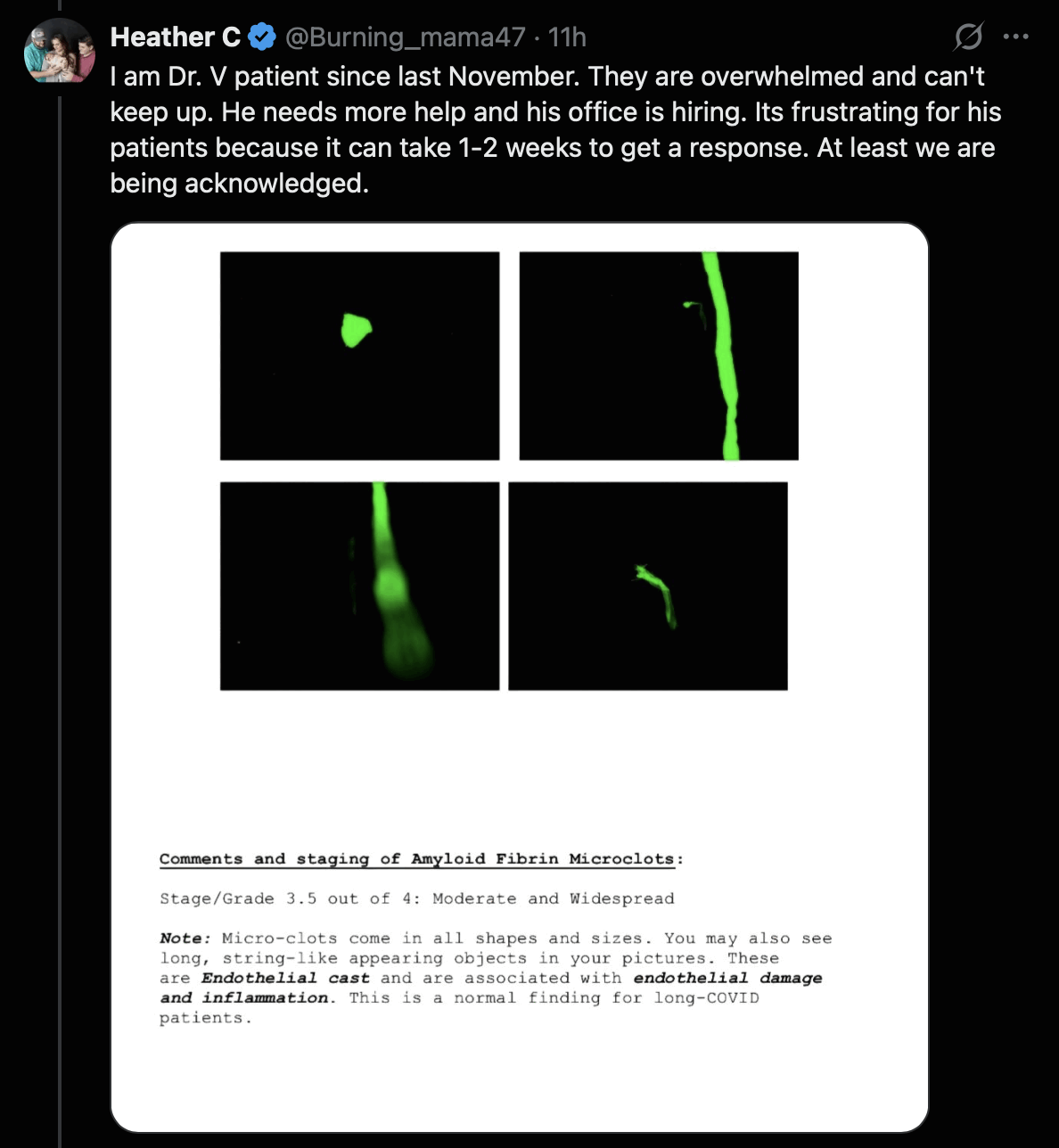
Jordan Vaughn in Alabama offers a similar services of testing for microclots as McCairn. One of his patients posted this tweet that showed a report they received from Vaughn, which said: "Micro-clots come in all shapes and sizes. You may also see long, string-like appearing objects in your pictures. These are Endothelial cast and are associated with endothelial damage and inflammation. This is a normal finding for long-COVID patients." The report included three images of elongated clots, even though none of them looked flat, and all had a fairly variable diameter, so they didn't look as much like textile fibers as some of the fibers that McCairn claims are microclots: [https://
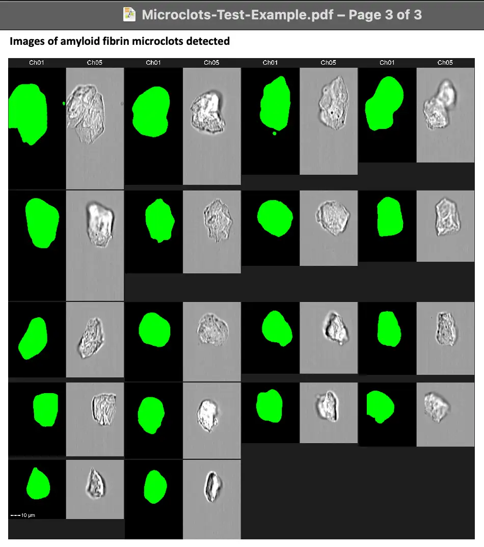
Apheresis Center in Cyprus also offers testing for microclots associated with long COVID. The website of the center includes these images of microclots, which all have a globular and not elongated shape: [https://
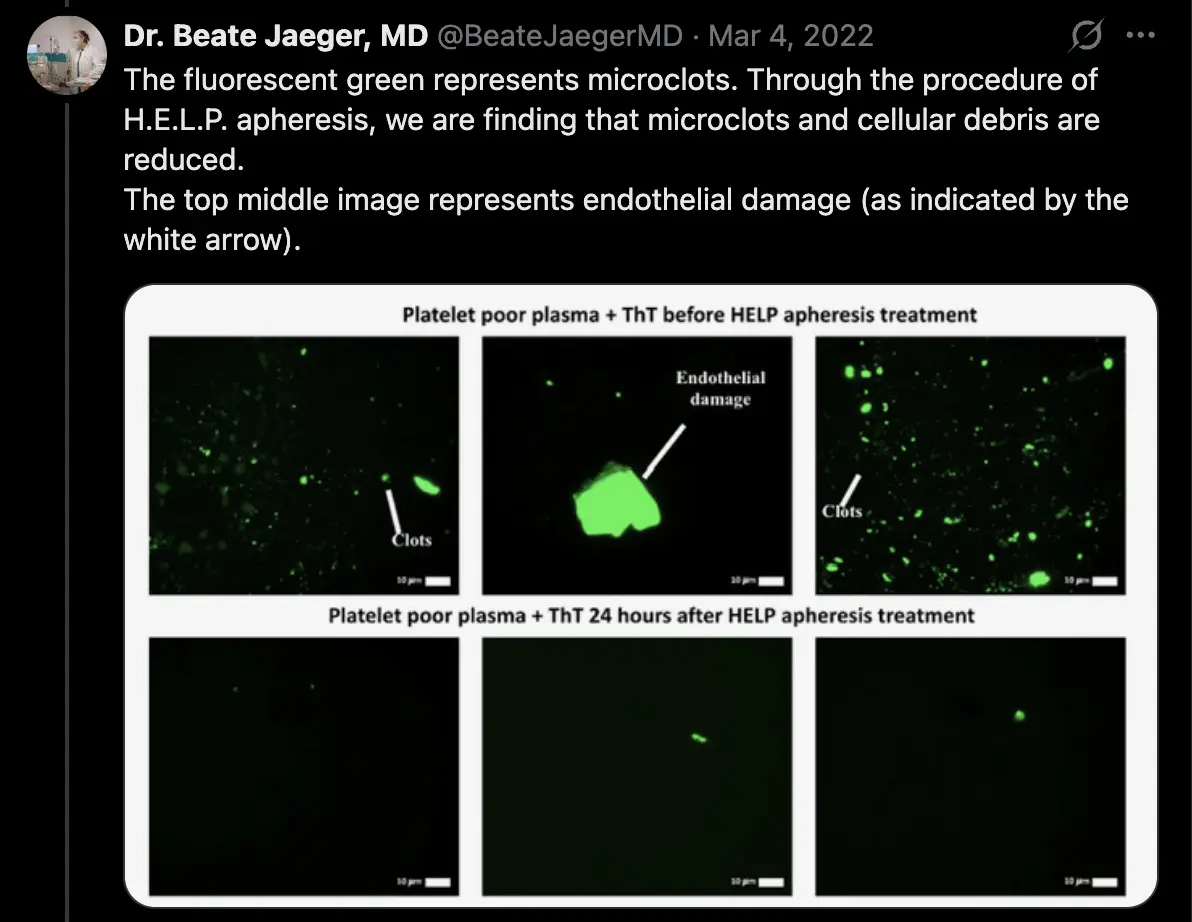
Beate Jaeger provides HELP apheresis treatment for long COVID in Germany. She posted these photos of microclots, which again have a globular or blob-like shape, and not an elongated shape: [https://
However in a paper by Jaeger from 2023 panel A3 shows a very long string-like structure with roughly uniform diameter: [https://
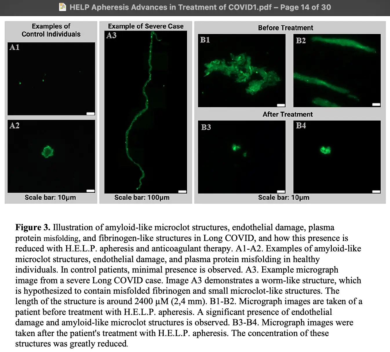
If the scale bar is correct, then the diameter of the string-like structure is around 20-30 µm, which would be too big to pass through the capillaries. Similarly the structures in panels B1 and B2 (labeled "before treatment") would be too big to pass through the capillaries. So I don't know if the structures are some kind of contaminants or artifacts. Jaeger's group doesn't seem to have verified if the string-like structure was actually made of fibrin, but they just said that it was "hypothesized to contain misfolded fibrinogen and small microclot-like structures".
Peter McCullough has developed a con where he sells a kit for testing the level of antibodies to the spike protein, and then he tells people with a high level of antibodies to buy his products in order to "detox" the spike protein, as if a high antibody level would be a sign that people have spike protein left over in their body. However in reality the presence of antibodies for the spike protein is generally a sign that immunity to COVID has been triggered by a vaccine or a natural infection. McCairn replied to a tweet about McCullough's test by saying: "Our patients show significant drops once treatment starts, and extends out for all tested patients. It's an indirect measure of the underlying pathology though." [https://
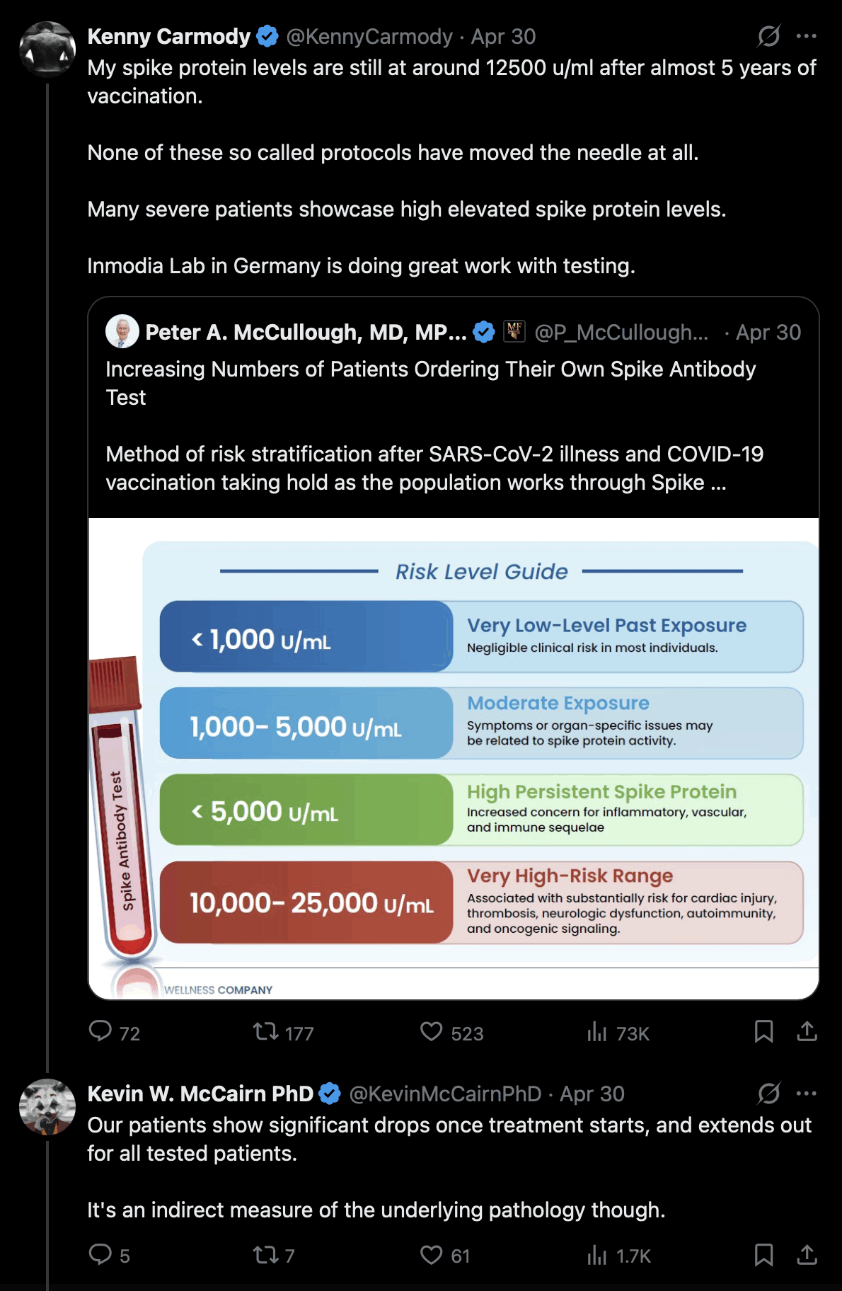
But how is it a symptom of a "pathology" if someone has antibodies for the spike protein? If McCairn's plasmapheresis treatment actually resulted in the permanent removal of spike antibodies, it might make his patients more susceptible to COVID, which would seem like a downside of the treatment. But of course the immune system will replenish the antibodies soon after the treatment. Antibodies are produced by memory B cells, which mainly reside in the lymph nodes, spleen, tonsils, and bone marrow. And even though a small part of memory B cells are circulating in blood, memory B cells that are removed from the blood still get replenished through a mechanism where the cells make copies of themselves through cell division.
One of McCairn's patients said that he was taking McCullough's Spike Detox supplements in order to reduce his level of spike protein antibodies, as if the antibodies would've somehow been harmful, or as if the anticoagulants in the Spike Detox supplements would've reduced the level of the antibodies. [https://
One of McCairn's patients posted the images below as evidence that her treatment was successful, because her blood looked more "stringy" before the treatment but more "smooth" after the treatment: [https://
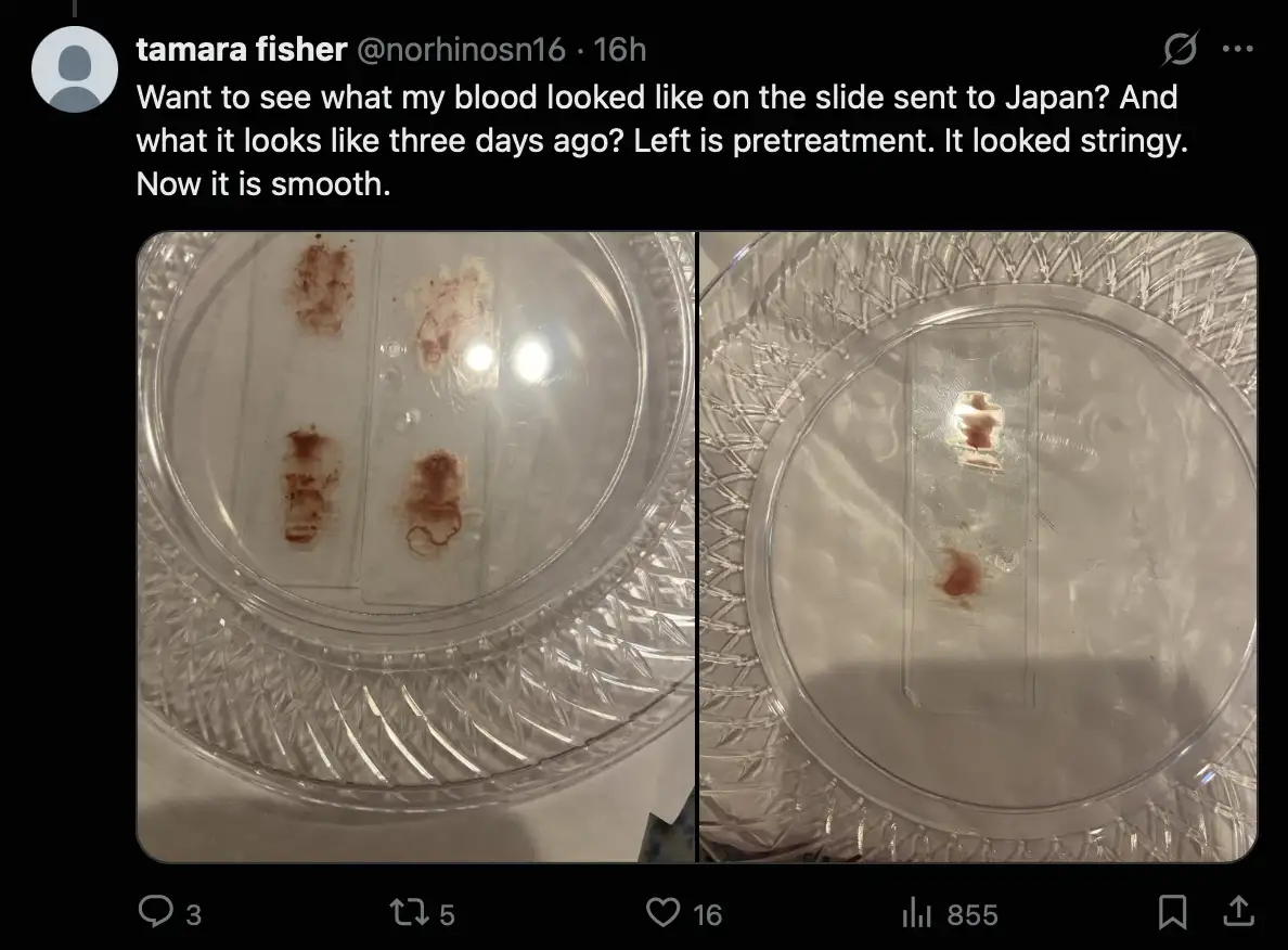
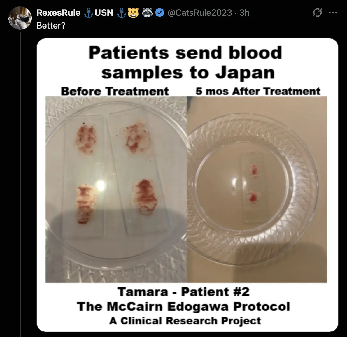
Her images were turned into an infomercial-style image by the memetic warrior from naval intelligence: [https://
A Twitter user whose display name is "china Victims of Sinovac COVID-19 Vaccine" was inspired by McCairn to get DFPP treatment in China. But he said "DFPP is completely ineffective; everything rebounded completely after four days. What a tragedy": [https://

He also said "DFPP has left my body weakened, and it delivered zero therapeutic effect". He was confused about whether he should've gotten DFPA or DFPP, but it's likely the results of DFPA wouldn't have been much different: [https://
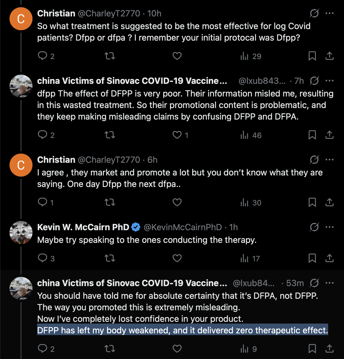
McCairn seems to have blamed the lack of success on getting DFPP and not DFPA or the baby tooth extracts. The Chinese guy was told by Shoujirou Katou that DFPP should be fine: [https://

Despite what McCairn said in the last tweet above, the Chinese user didn't accuse McCairn of malpractice, but only of not replying to his DM and not making clear enough if his protocol used DFPP or DFPA, and McCairn later admitted he wrong that the Chinese user hadn't sent a DM to him. [https://
Another Chinese user responded that RexesRule and CoyoteSanctuary also spoke about DFPP and not DFPA, and the Chinese user thought they were part of McCairn's marketing team: [https://

McCairn's patient mycityapartment wrote about the treatment that "I have experienced significant improvement that anticoagulants did not/could not provide". [https://
When I asked two of McCairn's patients if they had a rebound in symptoms similar to the Chinese guy, the patient mycityapartment said: "No I'm slowly improving across all symptoms including and especially neuropathy. 6 weeks from when I got home." [https://
But anyway, it was understandable for the Sinovac victims guy to mix up DFPP and DFPA, because even a promotional website about the treatment protocol says: "Edogawa Hospital's program is presented around Dual Filtration Plasmapheresis, also described as DFPP or DFPA in patient materials". [https://
It's also confusing how McCairn defines the acronym DFPA as "double filtration plasma apheresis", which is basically the same as the meaning of the acronym DFPP but with "plasma apheresis" written as two words and not in the more common form of "plasmapheresis" as a single word. So the similarity of the terms gives the impression that the term DFPA is a synonym of DFPP.
A Japanese guy who did McCairn's blood test said that the elongated fluorescent structure below was in his blood. The structure is fairly short, it has a variable diameter, it doesn't look too flat, and it doesn't have sharp folds, so it might be more likely to be actually made of fibrin than the typical fibers that McCairn finds in blood samples: [https://
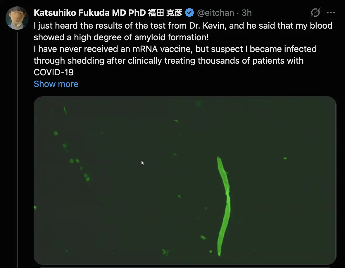
But McCairn still doesn't seem to be doing any procedure to determine the composition of the fluorescent structures, like immunostaining for fibrin antibodies or any kind of spectroscopy.
One of McCairn's patients from May 2026 said that he didn't notice any improvement after the plasmapheresis treatment: [https://
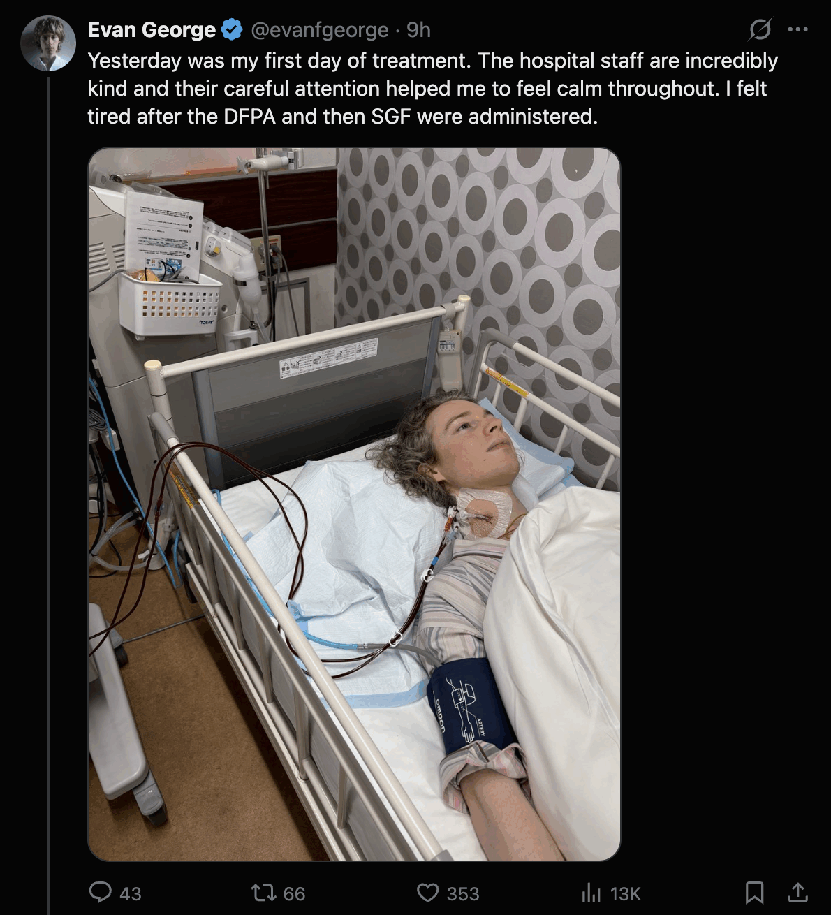

A few days later when he was asked about the effect of the baby tooth injections, he said: "It's subtle for me, but I definitely have been just a little bit more functional. I'm able to do a bit more before feeling sore but I still will start to feel symptoms if I push." [https://
In August 2026 Evan George posted another update, where he wrote: [https://
What is clear to me is that I began feeling better almost immediately after June and I arrived in Japan, before I had started any treatment. I don't know why. But it was apparent both in my walking data and in a general increase in energy and reduction in neuropathy. Even before beginning treatment, I was already walking more than I had been able to in the previous three years.
Once treatment began, however, it became increasingly difficult to separate those initial improvements from the effects of the treatment itself. Over the course of our time in Japan, I gradually went from walking around 5,000 steps a day to consistently reaching around 10,000 steps, often day after day without crashing. I could come home after walking that much and remain on my feet without the severe leg and joint pain that would previously have made that impossible.
One potentially important variable is the environment. I live in a relatively cold part of San Francisco that gets very little direct sunlight, whereas it was around 80°F when we arrived in Japan and became progressively warmer over the course of our stay.
Altogether, I was able to sustain this increased level of walking for roughly three months in Japan. During that period, I underwent four weeks of DFPA and five weeks of SGF, with the treatments spread out somewhat differently from the usual schedule because June has family in Tokyo. Without getting into all the subtleties of what did and didn't improve, the clearest changes were a significant reduction in my neuropathy, primarily in my legs and lower back and also in much of the severe cramping throughout my upper torso and into my lower back. Those symptoms improved quite quickly, and I was able to build on those gains while I remained in Japan.
Then I returned to San Francisco on August 5th. I was a dealing with a fever and respiratory illness in the context of my deficient immune function. Since returning home, my ability to walk and function has been progressively decreasing, while my neuropathy has been increasing again.
I'm still trying to understand why. At this point, I genuinely don't know how much of the improvement I experienced should be attributed to the treatment, how much may have been related to the environment in Japan, or what is responsible for the deterioration since returning home. I don't want to force a conclusion when I don't yet have one. All I can say with certainty is what I experienced: I improved substantially while in Japan, that improvement began before treatment, continued while I was undergoing treatment, and has not held since returning to San Francisco.
I informed Resia Pretorius of the experiment where McCairn mixed fibrinogen with ThT, shook the mixture, and claimed to have manifested a fibrin clot in the mixture (even though the fiber in his mixture looked like a dust fiber). She then quoted my tweet and wrote: "We and others looking for fibrinaloid microclot complexes, always do analysis on fresh blood plasma samples or plasma spun and stored at -80. We do not support dried sample analysis and any shipment at room temp. One must also be careful, as tissue paper fibers can show autofluorescence. However, we cannot comment on this exact photo." [https://
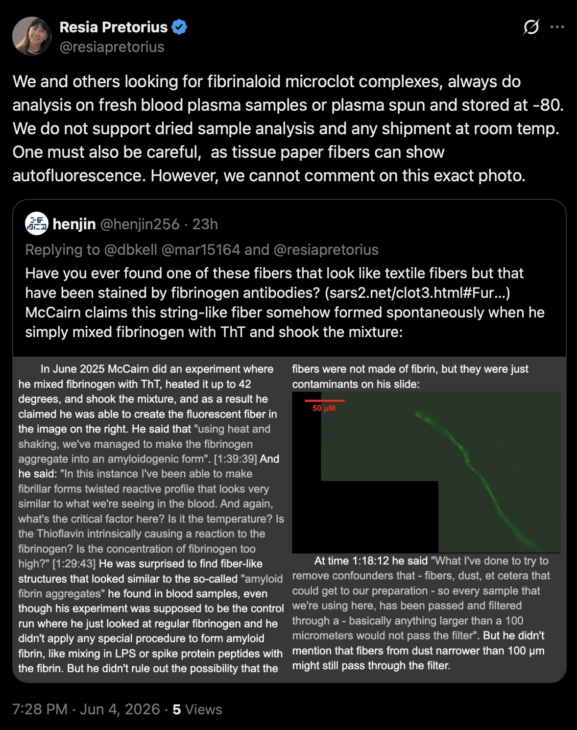
Grok said: "Tissue paper fibers fluoresce under UV light primarily due to optical brighteners (also called fluorescent whitening agents or FWAs) added during manufacturing. These colorless compounds, often stilbene derivatives or distyrylbiphenyl types, absorb invisible ultraviolet radiation (typically 340-370 nm) and re-emit it as visible blue light (around 400-450 nm) through fluorescence." Similar brighteners are also used in white textiles.
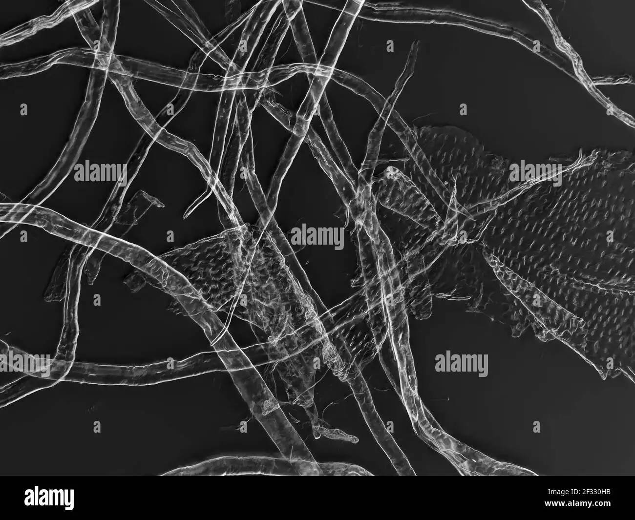
Tissue paper fibers look somewhat similar to cotton fibers and other cellulose fibers: [https://
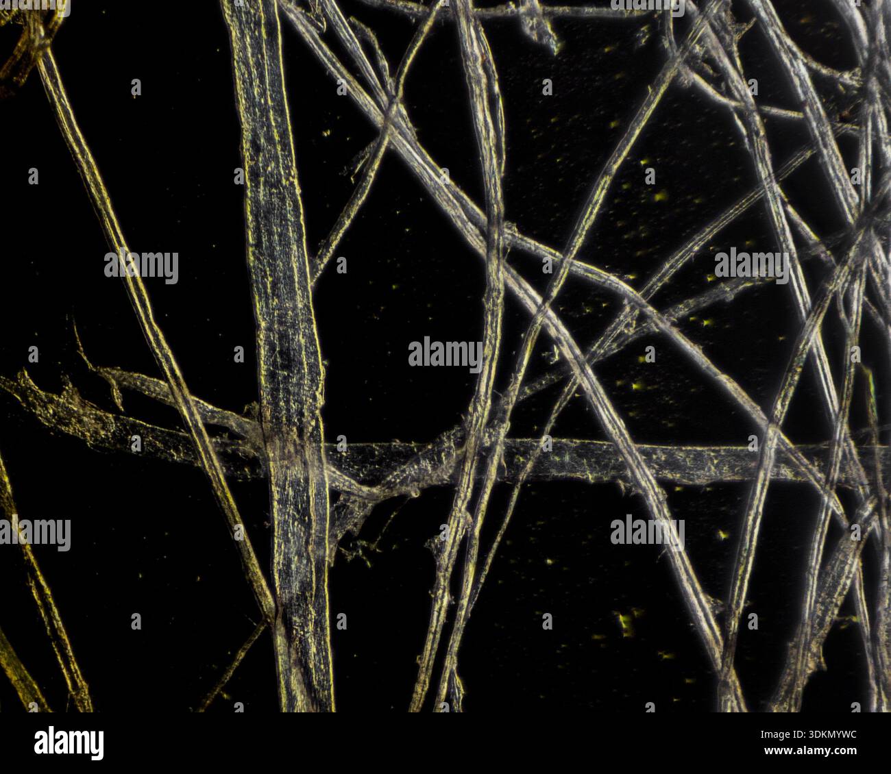
This shows toilet paper fibers: [https://
Resia Pretorius also posted these answers to replies to her tweet:

In June 2026 Mary Talley Bowden interviewed McCairn and his patient Ken Evans, whose username on Twitter is KenCaptn20114. [https://
At time 13:33 Ken Evans said: "We already know that we have spike proteins, we already know we have amyloid. We're just trying to survive. And that's the part of this whole Japan thing. I was not prepared for seeing how ill these people were. If you have a heart and you see these people, it will make you so mad that somebody is in this condition. And we all know that we had amyloids. They were stuck in all our tissues. And in my body, they were opening up in the sores. I was sneezing blood, I was coughing blood, blood in my feces, my stomach, my esophagus, liver, lungs, kidney, fibrosis, my swollen spleen, my heart. Every part of my body was absolutely devastated."
But how did he know he had "amyloids" in other tissues besides his blood? As far as I know, McCairn only tests for the presence of the amyloid fibrin clots in blood, and it wouldn't make sense for the fibrin clots to even exist in other tissues.
Ken Evans also seems to think that because he still has antibodies to the spike protein, it means that his body is full of spike protein coming from the vaccines. At time 16:58 he said that after he got McCairn's treatment, "My spike proteins went down 25%. So the treatment for me not only saved my life, but it pulled a lot of autoantibodies, out misfolding proteins, spike proteins, and all of those blood clots." So he seemed to think the reason why his level of spike protein antibodies was reduced by 25% was because he still had spike protein from the vaccines circulating in his blood but the spike protein was removed by McCairn's treatment, which doesn't make any sense.
At time 47:49 Bowden asked: "And just to be clear, normally in a normal blood you would not see anything lighting up at all?" Then McCairn showed the image below and said: "I mean you can see something. So look, if you look at that image that's on the screen, you can see smaller - like tiny microdots - that cluster around the image. Every sample will have that - because as the protein dries and desiccates on the slide, and there are some proteins and molecular species that are just fluorescent in and of themselves - but the size differential is what makes them different, okay. The fibrin causes large amyloidogenic twisting structures." Earlier McCairn has given the impression that the smaller fluorescent blobs are also be made of "amyloid fibrin", but now he seemed to suggest that the small blobs are not necessarily made of fibrin, but a characteristic of fibrin aggregates would be a large size or a twisting shape.

At time 1:26:04 Ken said: "People that are taking the vaccine is safe trail are ignoring Pfizer's own statistics. They admitted their vaccine didn't work. They admitted that it killed 82% of the pregnant women in studies."
But he misunderstood a set of preliminary data from the CDC's V-safe study, where at a time when almost no participants had yet completed a full-term pregnancy and only about 4% of pregnancies had completed, 104 out of 127 completed pregnancies had ended in a miscarriage (and not in the death of the pregnant woman). Later after more pregnancies had completed, the ratio of miscarriages divided by completed pregnancies dropped to about 13%. [https://
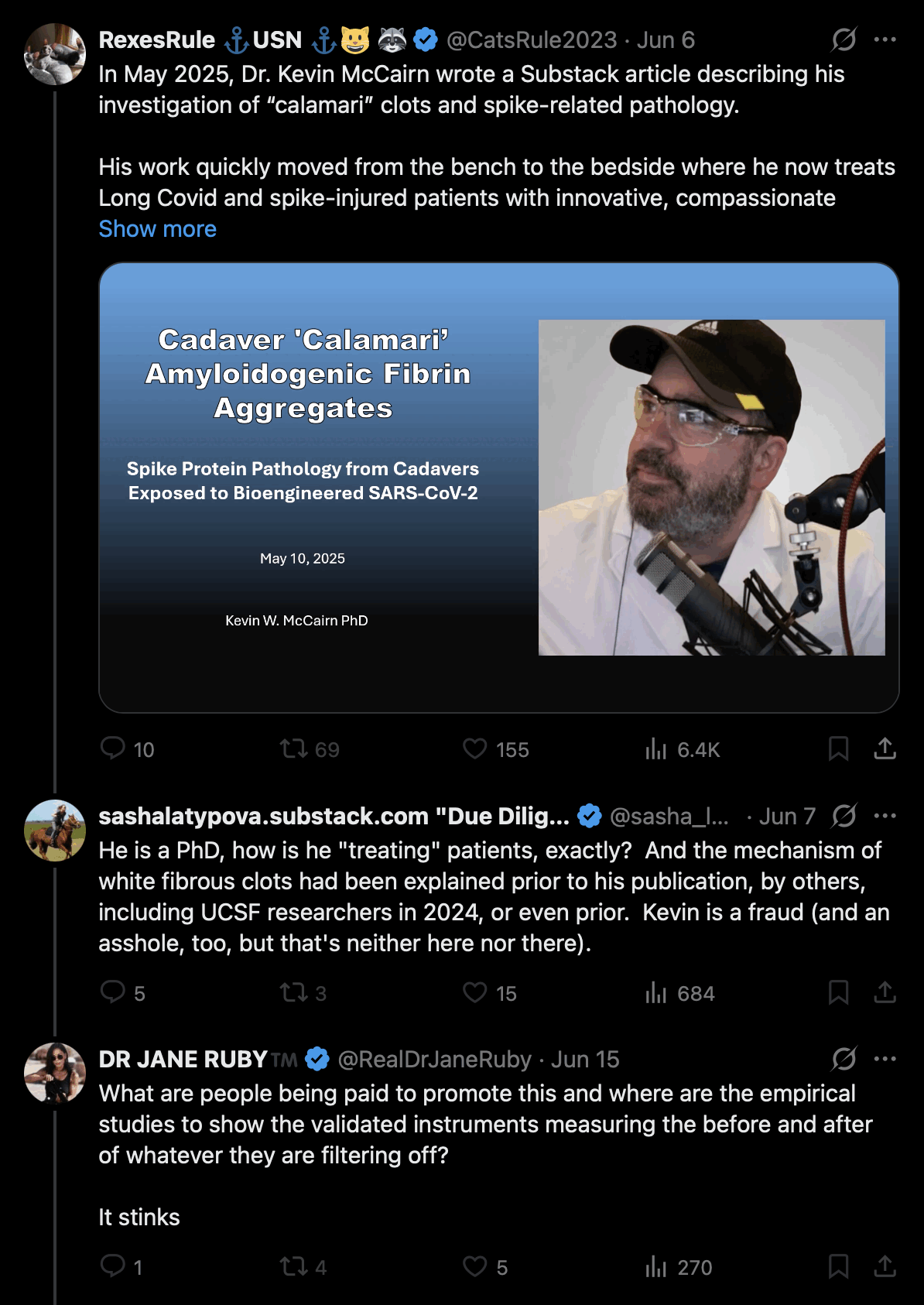
Now even Jane Ruby seems to think that RexesRule is being paid by McCairn to promote his treatment. Jane Ruby also asked "where are the empirical studies to show the validated instruments measuring the before and after of whatever they are filtering off", which is a very reasonable question: [https://
The plasmapheresis treatment at Edogawa Hospital is overseen by Shoujirou Katou, who is the director of the hospital. He also organized a conference at the hospital where the speakers consisted of Japanese anti-vaxxers and alternative medical practicioners: [https://

Katou's pinned tweet says in Japanese that the COVID vaccine is "extremely harmful and useless": [https://

The reason why Katou became the director of the hospital is not necessarily because of medical competency, but because the hospital was founded by his great-great-grandfather. In Japan it's common for the directorship of a hospital to be passed down from father to son. The previous director of the hospital was Katou's older brother.
One of McCairn's patients wrote: "Japan has the highest per capita mRNA vaccination rate in the world! According to Dr. Kato, 3.M Japanese died in the year following the initial rollout." [https://

In the years 2021-2024, there were about 1.5 million people per year who died in Japan, and virtually all of the people had eaten rice within a year from their death, so there were about 6 million Japanese people who died within a year from eating rice. But that doesn't mean that 6 million people were killed by rice, and in the same way the FOI data doesn't show that 3.89 million people were killed by vaccines. But the FOI data rather shows that vaccinated people have lower mortality than unvaccinated people.
The estimate of 3.89 million deaths was calculated based on a set of FOI data which included about 4% of the Japanese population, and which had about 150,000 deaths within a year from any vaccine dose, so the number of deaths was then multiplied by 25.8 to estimate the number of deaths in the whole Japan. [https://

Added in September 2026: Katou now posted this plot of the Japanese FOI data, where vaccinated people appear to have higher mortality rate than unvaccinated people: [https://

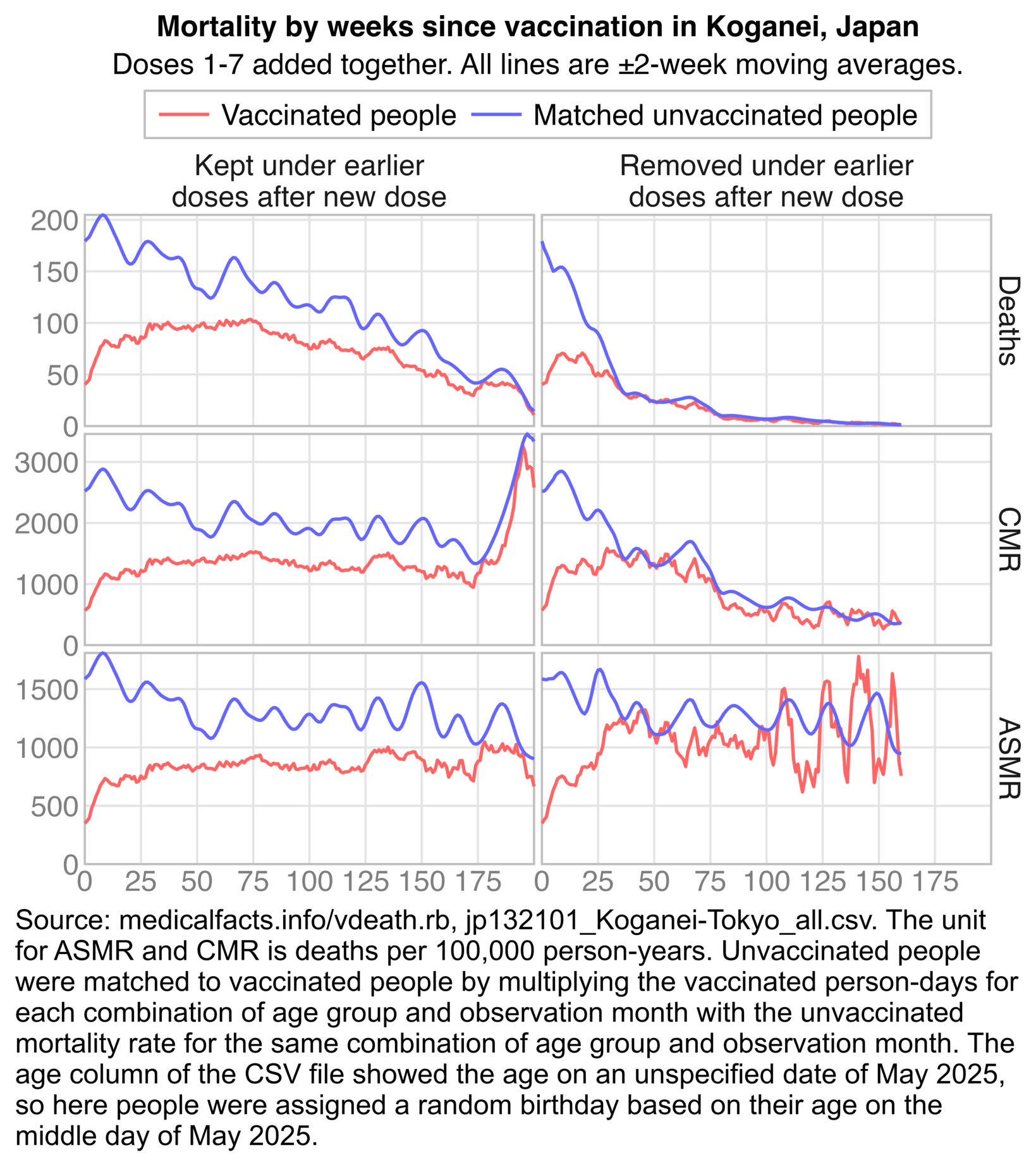
The plot has several problems, because it is supposed to show CMR but vaccinated people were older than unvaccinated people, so the plot should've been adjusted for age. Also vaccinated people with multiple doses were counted multiple times in the numerator for deaths, but only once in the denominator for population size. Also the dots for unvaccinated people show the number of deaths by weeks since March 8th 2021, and not so unvaccinated people have the same distribution of observation time as vaccinated people.
It would've been better to use the method I used in the next plot, where I matched unvaccinated people to vaccinated people by age group and observation month. My plot shows that after proper adjustment for age, observation month, and multiple doses, unvaccinated people have higher mortality than vaccinated people: [statistic2.
Now there's another cat lady who has started to say that McCairn is a hero who deserves a Nobel Prize. She made an AI-generated video where McCairn performs what looks like psychic surgery to "cast out amyloids": [https://

A Twitter user called Ben Liao posted the image below of a fiber in his blood found by McCairn. If McCairn's scale bar is correct, then the fiber has a diameter of about 10 µm: [https://

The fiber has a fairly uniform diameter which doesn't seem like a characteristic of a fibrin clot, but the fiber has the kind of a flat cupped body that is characteristic of cotton.
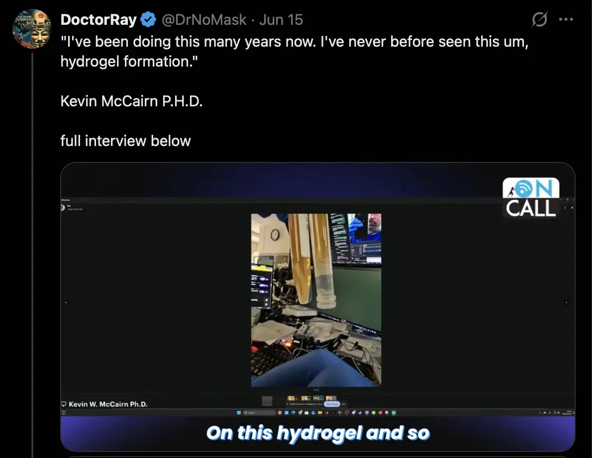
McCairn now claims that he is finding hydrogel in blood samples: [https://
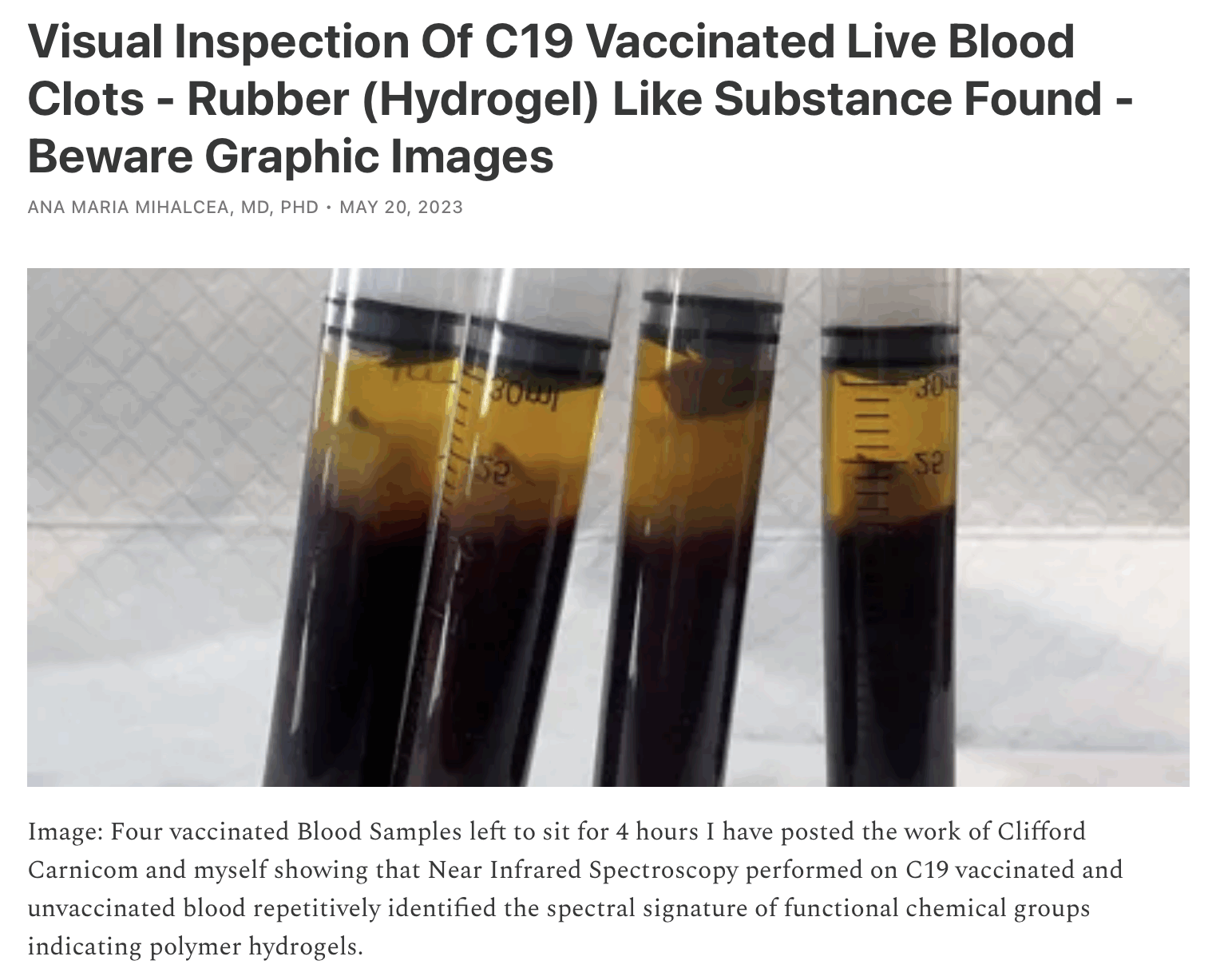
In 2023 Ana Mihalcea similarly said that she found hydrogel in vaccinated blood, and that the hydrogel was developing into calamari clots: [https://
McCairn is just rehashing old scams by Mihalcea. She also presented spectroscopy results of Hirschman's clots long before McCairn, and she said long before McCairn that a dust fiber in a blood sample was a precursor of a calamari clot in the process of being formed: [https://
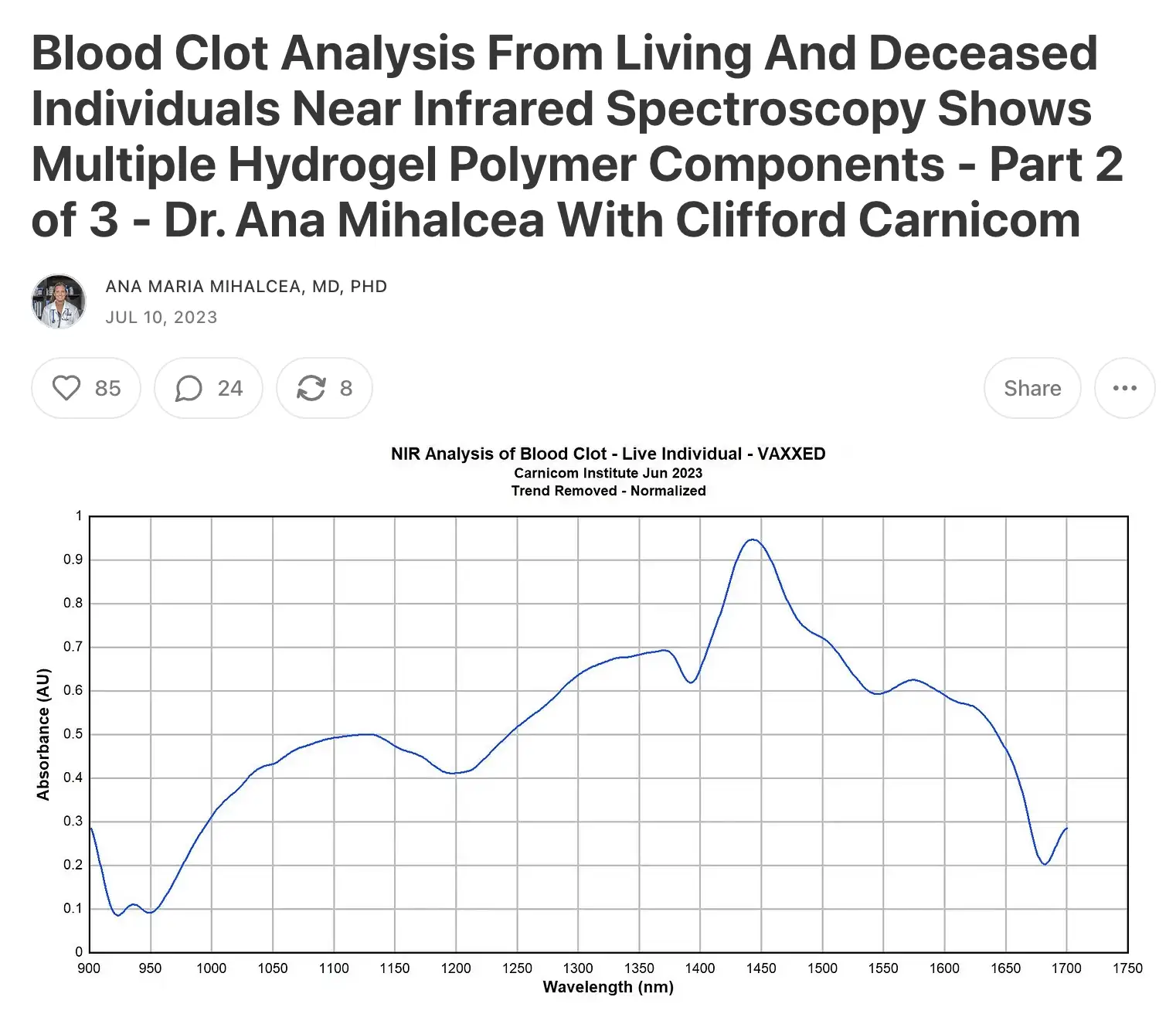
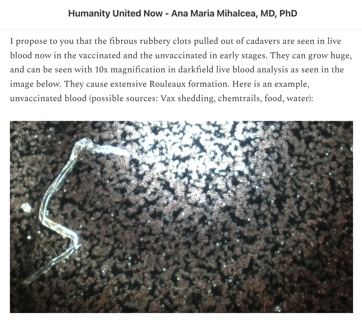
McCairn has been making fun of Mihalcea for a long time, and he calls her the "35,000-year-old cacodemon worshipper", because she is a member of a cult whose leader claims to be channeling 35,000-year-old Lemurian ascended master. But now McCairn himself is following in Mihalcea's footsteps, and he is trying to scare people into thinking that their blood contains mysterious hydrogel formations.
The story about the calamari clots was broken by Jane Ruby on the Stew Peters Network in January 2022. A few months earlier the Stew Peters Network broke Carrie Madej's story that vaccines contained hydras, and Carrie also told Stew that vaccinated people were growing blood clots that were made of hydras. In her debut video in July 2020, she said that COVID vaccines were going to contain DARPA hydrogel, which she said was a transhuman technology that would turn humans into cyborgs and hook up the human brain to the cloud. So long before she first presented the narrative about hydras, she was one of the primary people who helped popularize the narrative about DARPA hydrogel.
I believe the earliest person in alt media who started to say that COVID vaccines were going to contain DARPA hydrogel was Celeste Solum, who was already doing videos about DARPA hydrogel in March 2020, but she also said that COVID vaccines contained gorilla feces and neanderthal DNA: [https://


In July 2026 McCairn and some of his patients did a video with Mary Talley Bowden. [https://
At time 1:25:55, McCairn's patient Ken Evans said: "If people are listening, and they have symptoms like brain fog, massive headaches, neck aches, fatigue, shortness of breath, your vision - double vision, blurry vision - you get really dizzy, you can't stand up, your heart starts to race, or it starts to plummet, you get skin rashes, or bleeding, weird stomach issues, you're confused, you forget things a lot, you're - 'Oops, where was I?' - 'Oops, I just passed my exit while I'm driving' - severe muscle pain. These are all signs of microclots in your blood." And then he said people who have those symptoms should get their blood tested by McCairn (which will likely lead to McCairn finding microclots in the blood, and then suggesting people to get his 60,000 USD treatment).
Ken might have just as well listed the same litany of symptoms and then said they are a sign that you have Body Thetans, and the way to get rid of the Body Thetans is to pay 60,000 USD in auditing fees to McCairn. The list of symptoms Ken produced was so generic that everyone experiences nonzero of those symptoms to some extent. But I think neither McCairn or Ken can know if the symptoms have anything to do with microclots. And even if the plasmapheresis treatment would provide temporary relief to the symptoms, it doesn't necessarily have anything to do with removing microclots from the blood.
Scientologists believe that humans are possessed by disembodied souls of aliens called Body Thetans, and that symptoms of having Body Thetans include physical tightness or pressure in specific parts of the body, negative emotions like unhappiness, fear, or anger, or unexplained ailments, persistent fatigue, or cloudiness of thought. [https://
Ana Mihalcea wrote that the symptoms of Morgellons disease have a lot of overlap with the symptoms of vaccine injury and long COVID. She wrote: "There is a huge overlap between C19 vaccine injury, shedding and long Covid symptoms with the suffering of Morgellon's victims. In particular, chronic fatigue, brain fog, shortness of breath with mucus production, anxiety, blurry vision, ringing in ears, headaches, circulatory problems and palpitations, joint stiffness, digestive problems, strange sensations under the skin and more - all have significant overlap." [https://
Some lady on Twitter posted the image below, posted a link to her GiveSendGo page, and wrote: "The first picture is an amyloid macro clot (next size up from a micro clot) that Dr. McCairn found in my husband's blood sample. Just like my husband, my son and I (second picture) were told we also have severe micro clots. We need to get to Japan for relief and treatment with the McCairn Edogawa protocol - Dual Filtration Plasmapheresis (DFPP) and Stem Cell Growth Factors (SGF). These are some of our symptoms collectively: headaches, fatigue, trouble exercising, stomach cramps, nausea, chronic on/off vomiting, heart pounding, neuropathy, feeling hot/cold, bladder issues, eye and ear inflammation, AFib, sleeping problems, severe depression, brain fog, anxiety, memory issues, mast cell, and cytokine storm. Please share, pray, and give. Thank you!" [https://
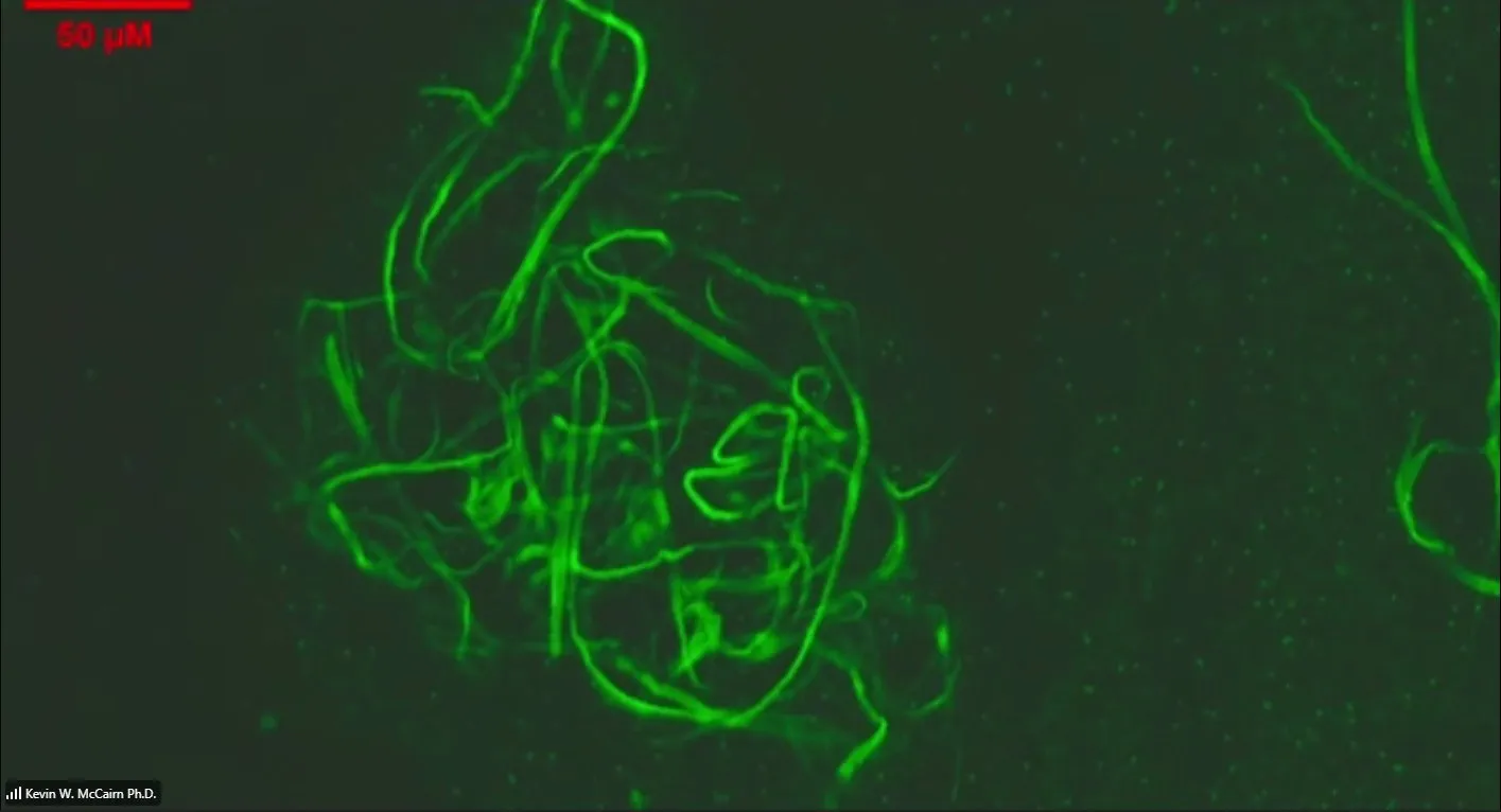
Just about anyone experiences the symptoms she listed to some extent, but McCairn seems to have convinced her that she now needs to get his expensive treatment because of the symptoms.
Resia Pretorius said that the image above "does not look like microclot complexes": [https://
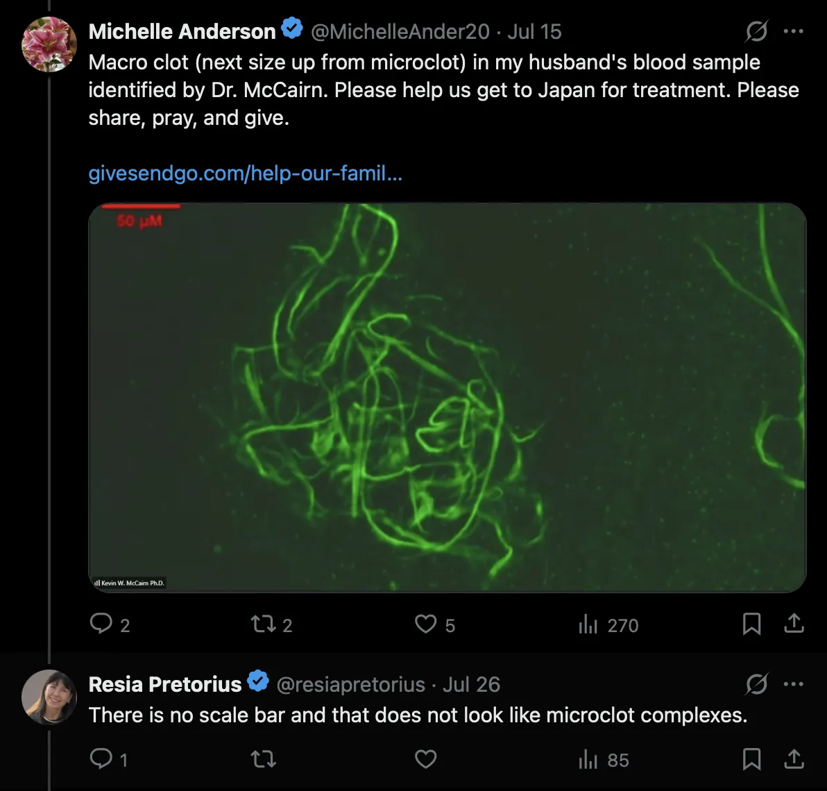
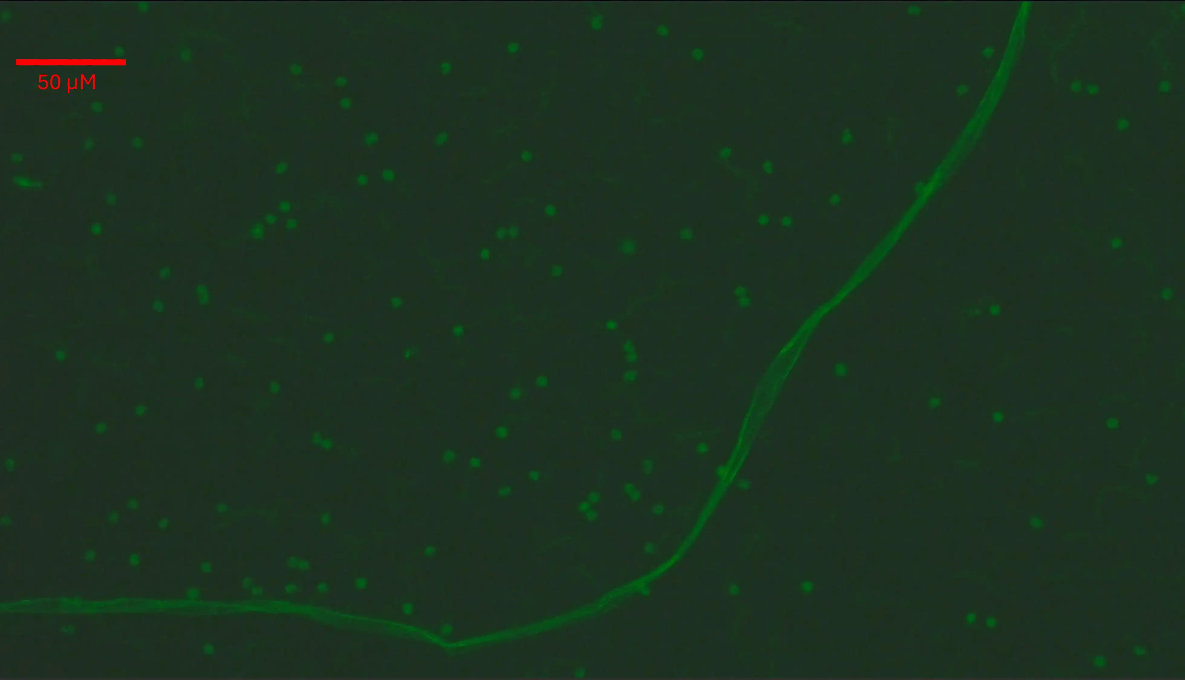
She said the image doesn't have a scale bar, but I guess she missed the scale bar in the top left corner (or maybe she was confused by how McCairn used micromolars and not micrometers as the units of his scale bar).
But anyway, I'm not sure if McCairn screwed up his scale bar yet again, because based on the size of both the fibers and the small green blobs, the image looks like it might be taken at around 5 times lower magnification than indicated by the scale bar. Based on the 50 µm scale bar, the small green blobs in the image would be only about 1 µm wide, even though in McCairn's other microscope images, similar green blobs have been about 5 µm wide (even though I'm not sure what the blobs are supposed to be, or if different images feature different types of blobs).
If the scale bar is actually around 250 µm and not 50 µm wide, then the diameter of the spaghetti-like fibers would fall around the typical range of 10-30 µm. But if the scale bar is actually only about 50 µm wide, then the fibers would be much narrower than typically.
Resia Pretorius also posted these tweets about McCairn's protocol: [https://



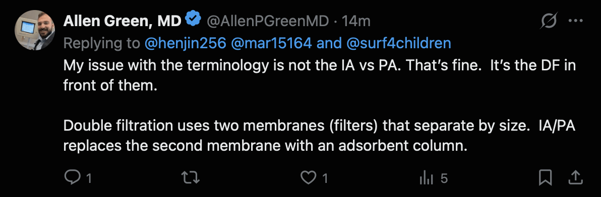
Allen Green is an associate medical director of a plasma apheresis center. He posted these tweets about McCairn's treatment protocol: [https://
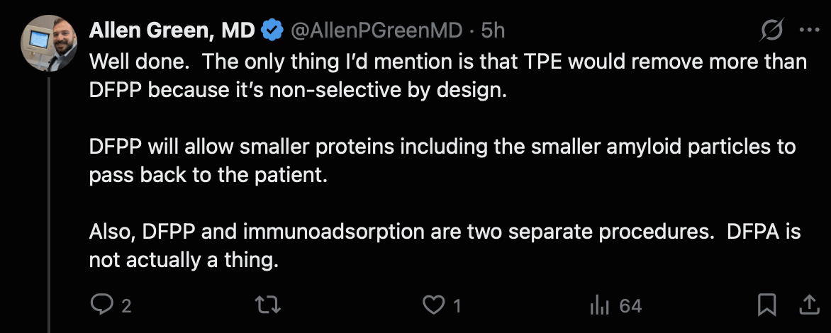



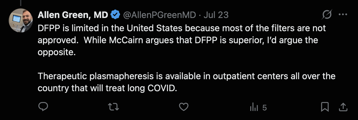

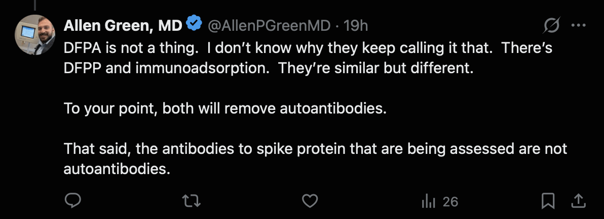

Allen Green said that "DFPA is not a thing". When I googled for "dual filtration plasma apheresis" in double quotes, there were only 7 pages of results, which all appeared to be about McCairn's protocol.
There's an RCT of therapeutic plasma exchange as a treatment for long COVID, which found no significant difference between the treatment group and placebo group. [https://


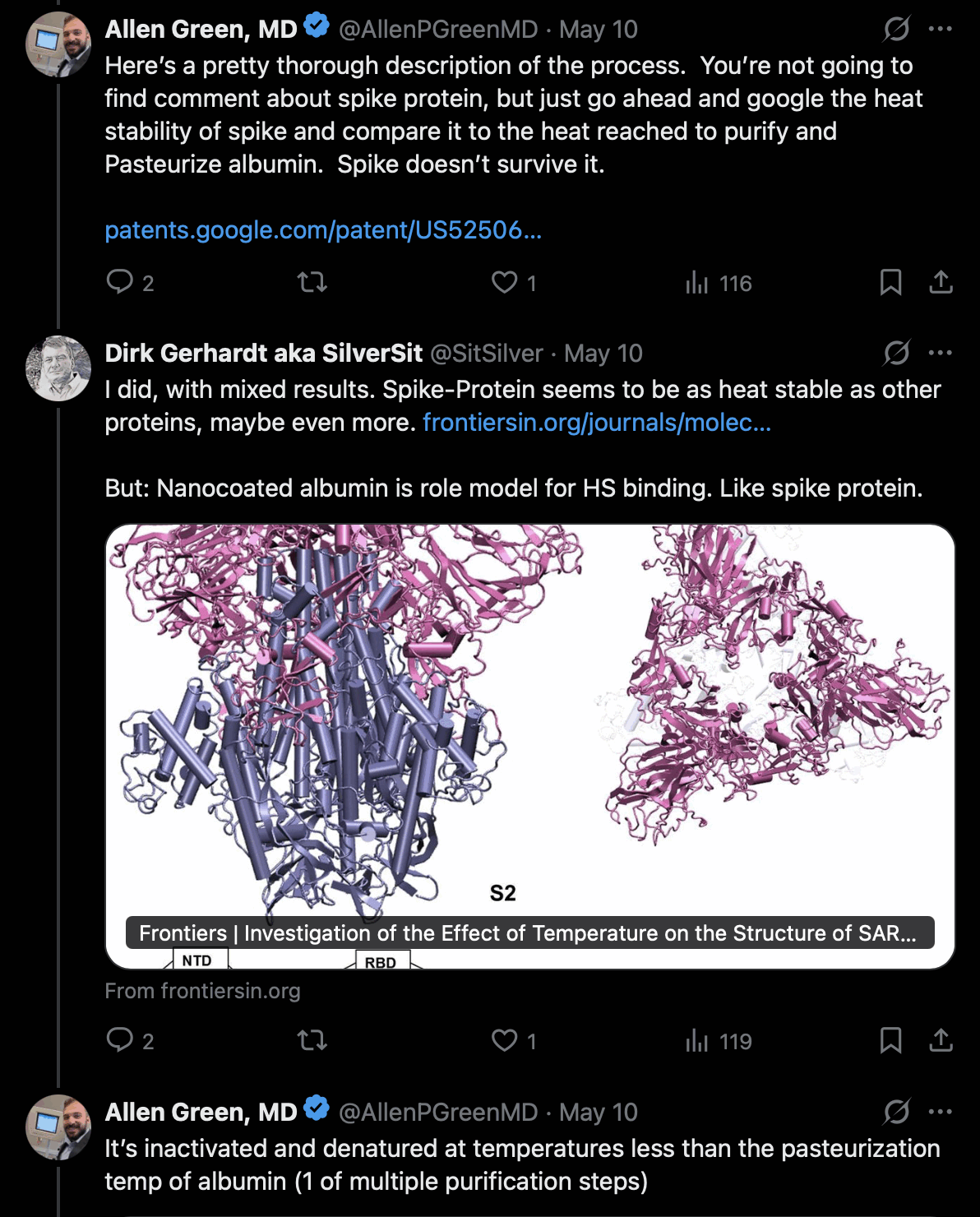
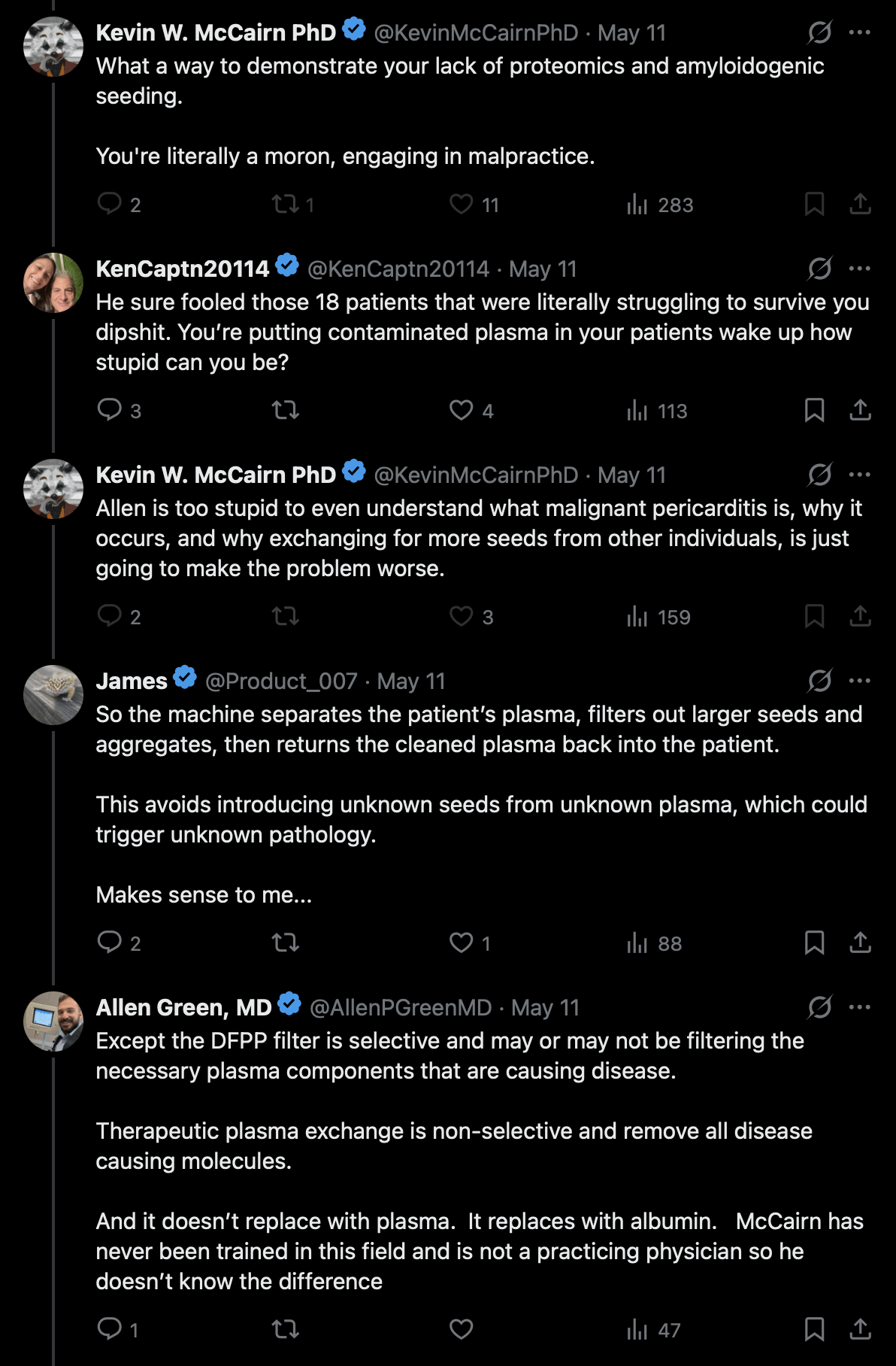

In the thread above McCairn referred to albumin as a "peptide", so he seems to have still not learned the difference between a peptide and a protein. But next he ironically said that Allen Green struggles with basic molecular biology.
McCairn has earlier also said that fibrin is a peptide:
In June 2026, The Lancet published a paper about a German RCT of immunoadsorption as a treatment for long COVID. The trial empolyed the same Asahi TR-350 immunoadsorption column that is used by the McCairn-Edogawa protocol. After 5 cycles of treatment given over the course of a single week, no significant benefit was found compared to a sham treatment (even though there were only 20 patients per arm, so the lack of significance may have been due to a small number of patients): [https://
Background
Post-Covid syndrome is a debilitating condition which may be caused and/or aggravated by autoantibodies. The current study aimed to determine whether autoantibody depletion by immunoadsorption is effective to reduce the symptom burden of patients with post-Covid syndrome.
Methods
IAMPOCO was a randomised, patient-blinded, sham-controlled crossover trial of immunoadsorption with tryptophan adsorbers versus sham treatment in patients with post-Covid syndrome at a tertiary academic care centre. The primary outcome was the difference in change in symptom severity before and after the respective therapies. Secondary outcomes included treatment safety, prevalence of autoantibodies against G protein-coupled receptors (adrenergic and muscarinergic receptors), and the influence of treatment on autoantibody levels.
Findings
40 patients with post-Covid syndrome and a symptom severity of at least 2 on the Post-Covid-19 Functional Scale were included and randomised to a treatment sequence. There was no difference in change in symptom severity between immunoadsorption and sham; odds ratio in Post-Covid-19 Functional Scale, OR = 1.17 (95% CI, 0.41-3.36; p = 0.771), mean difference in MFI-20 2.4 (95% CI, -3.7-8.5; p = 0.437), in CFS 0.09 (95% CI, -4.5 to 4.7; p = 0.970), in Bell-Scale -2.6 (95% CI, -6.9 to 1.8; p = 0.246), in MoCA score -0.01 (95% CI, -1.2 to 1.1, p = 0.993) and in Handgrip-strength deviation from individual normal value 1.3 (-0.83 to 3.5, p = 0.234). 34 adverse events occurred, 10 during or after sham treatment and 24 during or after immunoadsorption. Autoantibodies against G protein-coupled receptors were depleted by immunoadsorption but not sham treatment.
Interpretation
Immunoadsorption was not effective in reducing symptom burden in post-Covid syndrome.
Earlier when the Sinovac victims guy got DFPP in China but he said the treatment was ineffective, McCairn blamed it on the treatment being DFPP and not DFPA. It's not clear if DFPA is supposed to mean regular immunoadsorption, or if DFPA means DFPP combined with immunoadsorption. But in either case, the German trial suggests that the TR-350 immunoadsorption column is not necessarily effective in treating long COVID.
A placebo effect was evident in some patients who received a sham treatment in the German study, because the study said: "On the other hand, both of the 2 subjects that left the study after their initial sham treatment reported an improvement and stated that they did not want to risk receiving what they mistakenly thought would be sham treatment in the second cycle (when they would have been treated with true immunoadsorption)."
One of McCairn's patients described the treatment protocol like this: "My blood is drawn out through a catheter in my neck. The plasma is separated from my blood cells and run through a column called the IMMUSORBA TR-350, made by Asahi Kasei Medical in Japan. Inside that column, the pathological antibodies and immune complexes circulating in my blood bind directly to the column material and are captured. My own cleaned plasma is then returned to my body." [https://
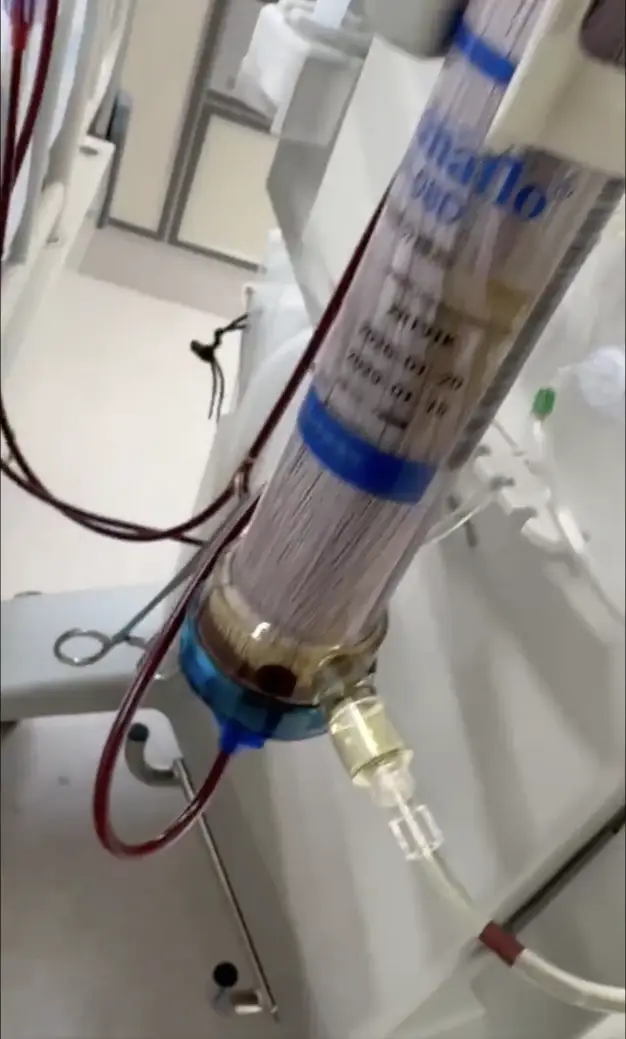
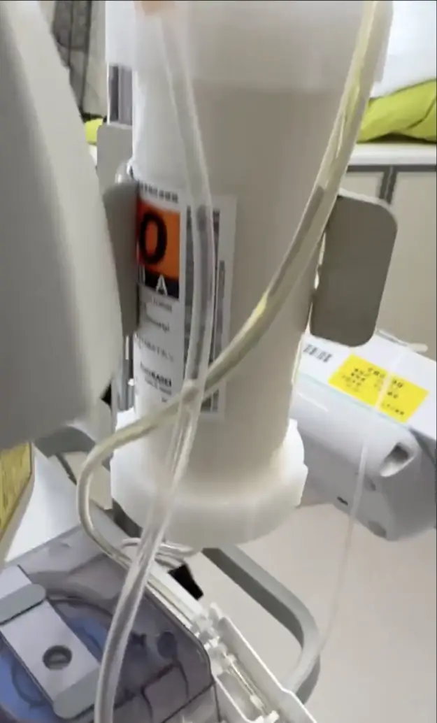
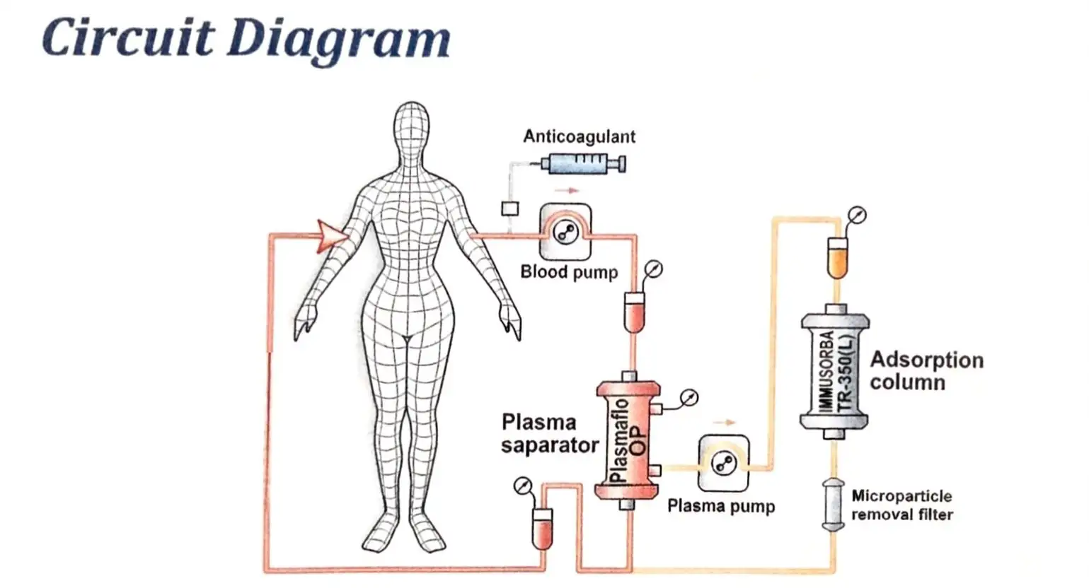
She also posted this circuit diagram for IMMUSORBA TR-350, which includes the plasma separator (that is common to both DFPP and immunoadsorption) and an immunoadsorption column, but the diagram is missing the plasma fractionator that acts as the second filter in DFPP: [https://
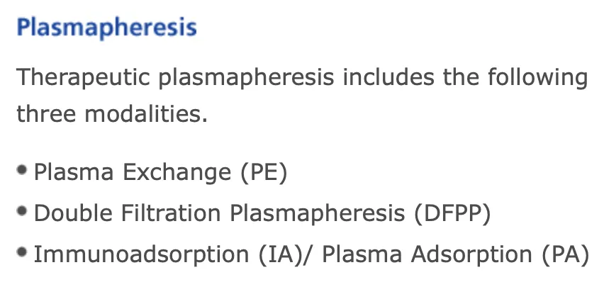
"Plasma adsorption" seems to be an alternate term for immunoadsorption that is mainly used in Japan. The Japanse manufacturer Asahi's website speaks of plasma adsorption interchangeably with immunoadsorption: [https://
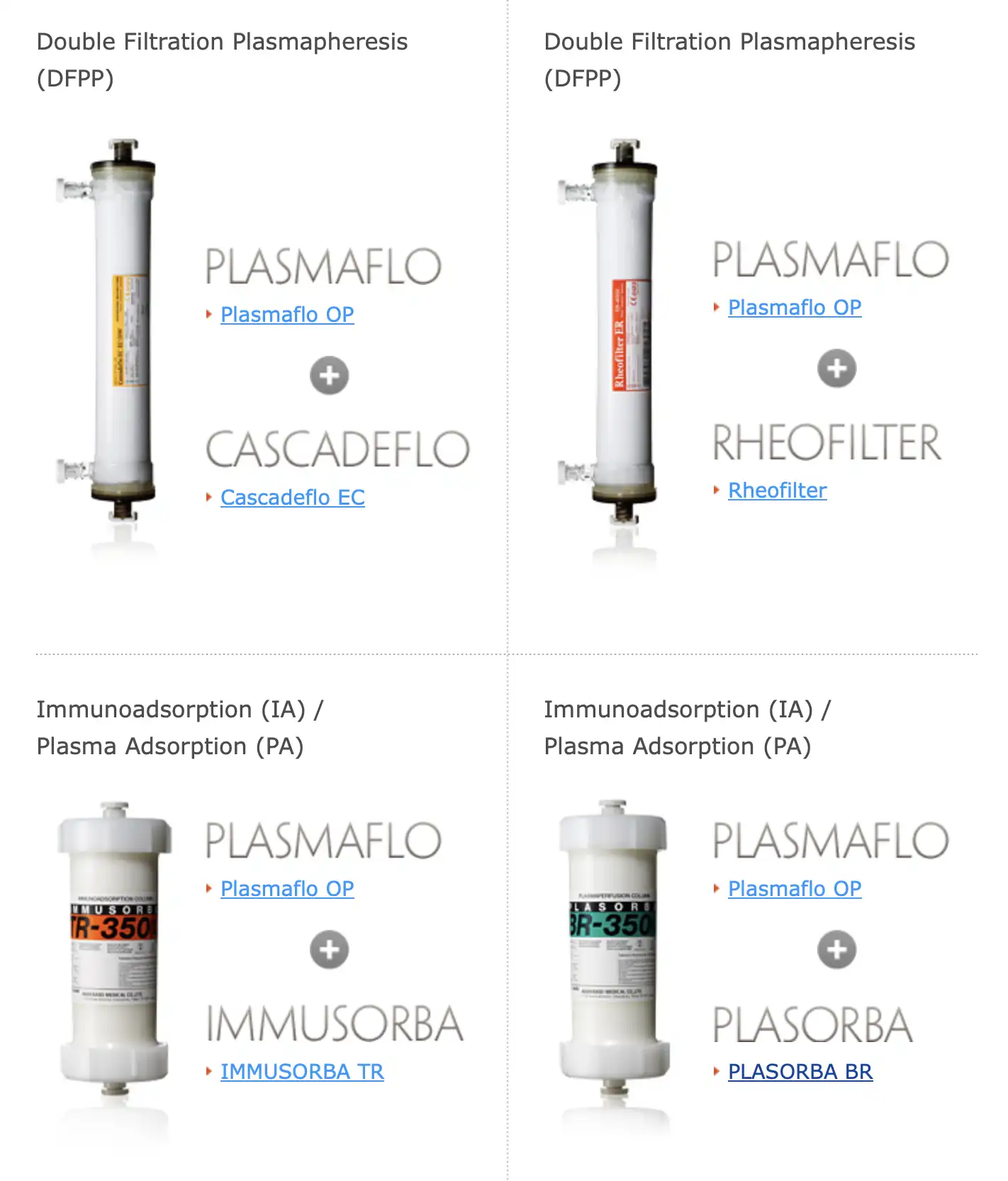
The setup shown in the video by McCairn's patient corresponds to the bottom left immunoadsorption setup here, and not to the DFPP setups on the top row:
I asked Shoujirou Katou on Twitter if what he calls DFPA is some kind of a hybrid between DFPP and IA, or if DFPA is just a nonstandard term for regular IA, but he didn't answer me. [https://
Grok said:
Immunoadsorption (IA) and Plasma Adsorption (PA) are very closely related and often used interchangeably. On Asahi Kasei's official site, they are grouped together as one modality: "Immunoadsorption (IA) / Plasma Adsorption (PA)". The main technique involves separating plasma and then running it through a selective adsorption column (such as the IMMUSORBA TR-350 tryptophan column) that binds specific pathogenic substances like antibodies and immune complexes. The cleaned plasma is then returned to the patient. This is distinct from Double Filtration Plasmapheresis (DFPP), which relies on two membrane filters for size-based separation rather than chemical adsorption. In the context of Edogawa Hospital, the treatment using the TR-350 column is referred to as Plasma Adsorption, and they brand the overall approach as DFPA.
The term "Plasma Adsorption" (PA) is used mainly by Asahi Kasei and in Japanese apheresis literature, though it is not entirely exclusive to them. Asahi Kasei officially lists it alongside immunoadsorption as one of the three main plasmapheresis modalities. In Japan it is a standard clinical term for column-based selective plasma purification. Internationally the term appears in some literature, but Western sources more commonly use "immunoadsorption" or "selective plasma adsorption." When you see "plasma adsorption" mentioned in relation to Edogawa Hospital or the McCairn protocol, it specifically refers to their use of Asahi's IMMUSORBA columns. Overall, it describes essentially the same column-based technique as immunoadsorption, with naming varying by region and manufacturer.
Allen Green pointed out that the term DFPA (where the DF stands for "double filtration" or "dual filtration") is a misnomer, because it implies the use of a second filter like in DFPP, even though the TR-350 column used at Edogawa is an adsorption column and not a filter: [https://
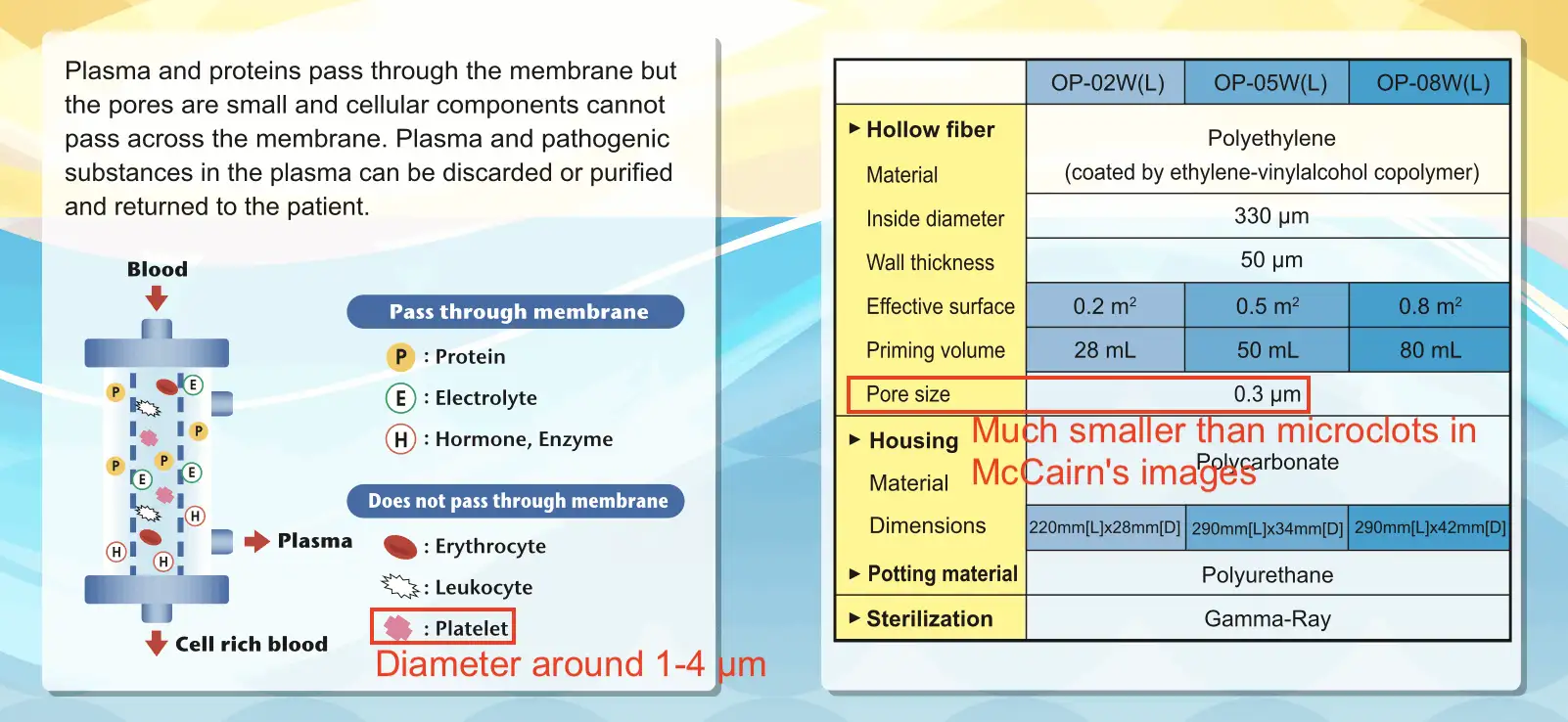
Asahi's PLASMAFLO plasma separators have a maximum pore size of 0.3 µm, so microclots that are too big to pass through the pores don't pass to the immunoadsorption column, but the clots are rather returned to the patient along with the red blood cells, so that the clots never get filtered. [https://
[Patient Blood] ──> [PLASMAFLO OP Filter (0. 3 µm)] │ │ ▼ (Through Pore) ▼ (Main Path) [Isolated Plasma] [Cell- Rich Blood] (No Microclots) (Contains 10- 30 µm Clots) │ │ ▼ │ [IMMUSORBA TR- 350] │ (Binds Antibodies) │ │ │ ▼ ▼ [Cleaned Plasma] ─────> [Recombined Blood] ──> [Return to Patient]
Asahi's brochure for their PLASMAFLO plasma separators shows that platelets are too big to pass through the pores, but platelets typically have a diameter around 1-4 µm: [https://
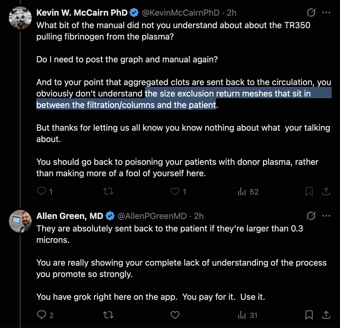
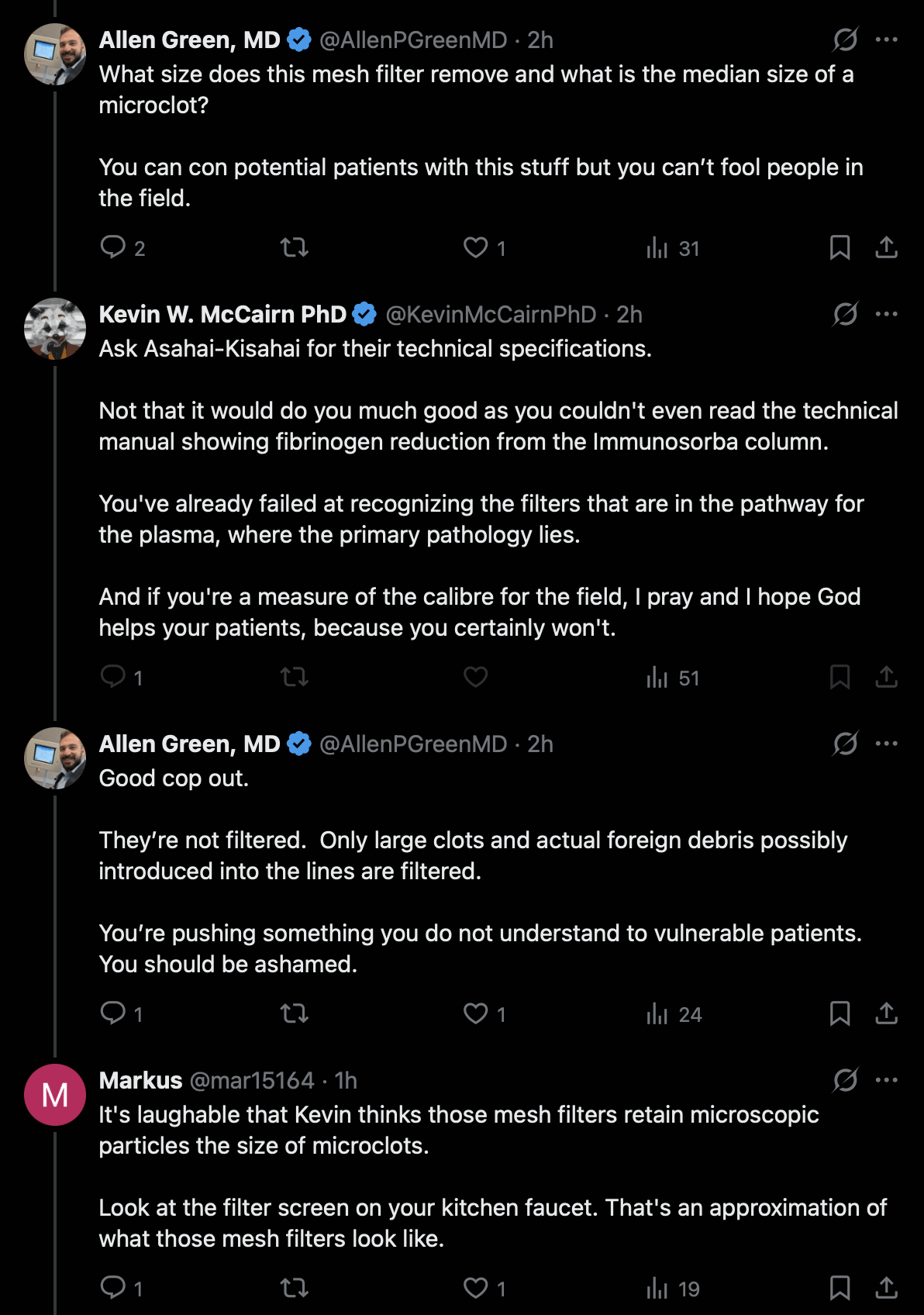
McCairn said that the IMMUSORBA column "seems particularly effective at clearing amyloidogenic fibrin". When Allen Green pointed out that the microclots are too big to pass through the pores to the IMMUSORBA column, McCairn said that was why there were two filters. So McCairn doesn't seem to understand that the microclots don't pass through the plasma separator filter, so the clots are never routed to the TR-350 column (which is not even a filter but an adsorption column): [https://

McCairn responded by saying there was a size exclusion mesh after the IA column, because he didn't seem to understand that the microclots don't get passed to the IA column in the first place: [https://
Then McCairn said that after the plasma is combined with the cell-rich-blood, there's a mesh filter before the combined blood is returned to the patient, but he didn't seem to realize that the holes in the mesh were far too large to filter out microclots: [https://
Google AI said this about the "venous line air trap mesh filter" that is located after the spot where the plasma gets combined with the cell-rich blood:
In an Asahi Kasei Medical apheresis circuit, the standard inline venous line blood chamber uses a mesh filter with a hole size typically ranging from 150 µm to 200 µm (micrometers).
The primary purpose of this standard blood mesh filter is to act as a gross macro-aggregate trap right before the blood safely returns to the patient's body.
Purpose of the Mesh Filter Holes
Traps Macro-Clots: It catches any blood clots, fibrin strands, or large particulate matter that may have formed due to blood pooling or surface activation within the extracorporeal tubing.
Catching Persistent Microbubbles: The mesh surface tensions help disrupt and break down macro-air bubbles, assisting the main bubble catcher chamber.
Low Fluid Resistance: Because whole blood cells (red blood cells are roughly 7 to 8 μm) must pass through this stage after recombining with the plasma, a large 150 to 200 μm opening ensures zero resistance, zero clotting of normal blood cells, and stable venous pressures.
So if we assume the microclots have a diameter between 1 and 100 µm, then the clots are too big to pass through the 0.3 µm pores in the plasma separator, so the clots never pass through the "size exclusion return mesh" that is located behind the immunoadsorption column but before the spot where the plasma gets combined with cell-rich blood. The "air trap mesh filter" is located after the spot where the plasma gets combined with cell-rich blood, but the pore size of the filter is around 150-200 µm, which is too large for the microclots to get trapped. So none of the three filters ends up removing the microclots from the patient's blood.
Now after McCairn had been made aware of a fatal flaw in his protocol, he didn't acknowledge the flaw, but he tried to change the topic by claiming that his protocol still reduces clots in the blood, because he showed the photo below and wrote: "And here just for you, pre-treatment hyper-coagulable plasma (LHS), note the large fibrin clot down the side of the tube, and post treatment (RHS)." [https://
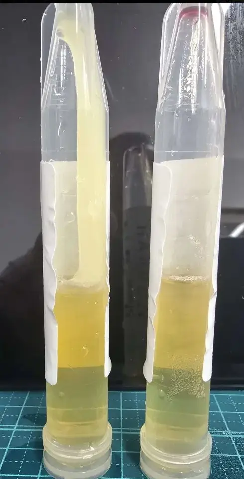
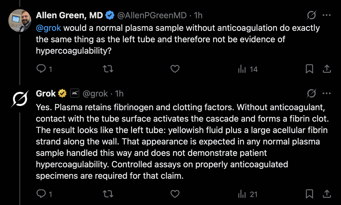
But Allen Green pointed out that it's normal for plasma to form clots in a tube: [https://

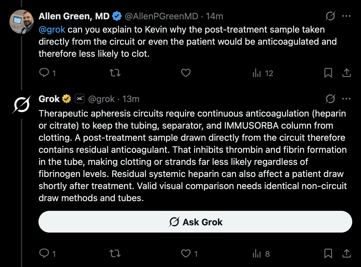
McCairn said that the plasma on the right side was taken out of the apheresis circuit, but Allen Green pointed out that anticoagulants are added to blood to prevent clotting in the apheresis circuit: [https://
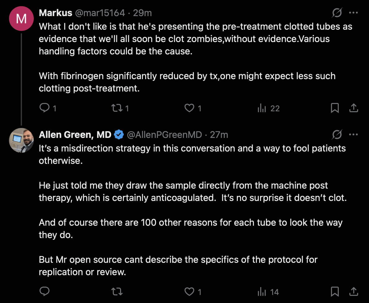
Allen Green also pointed out that IA treatment is designed to not remove too much fibrinogen, because it would increase bleeding risk, and the level of fibrinogen recovers after about 2-3 days anyway: [https://

There's two studies where IA treatment using TR-350 reduced the fibrinogen level in blood by about 60-70%. [https://
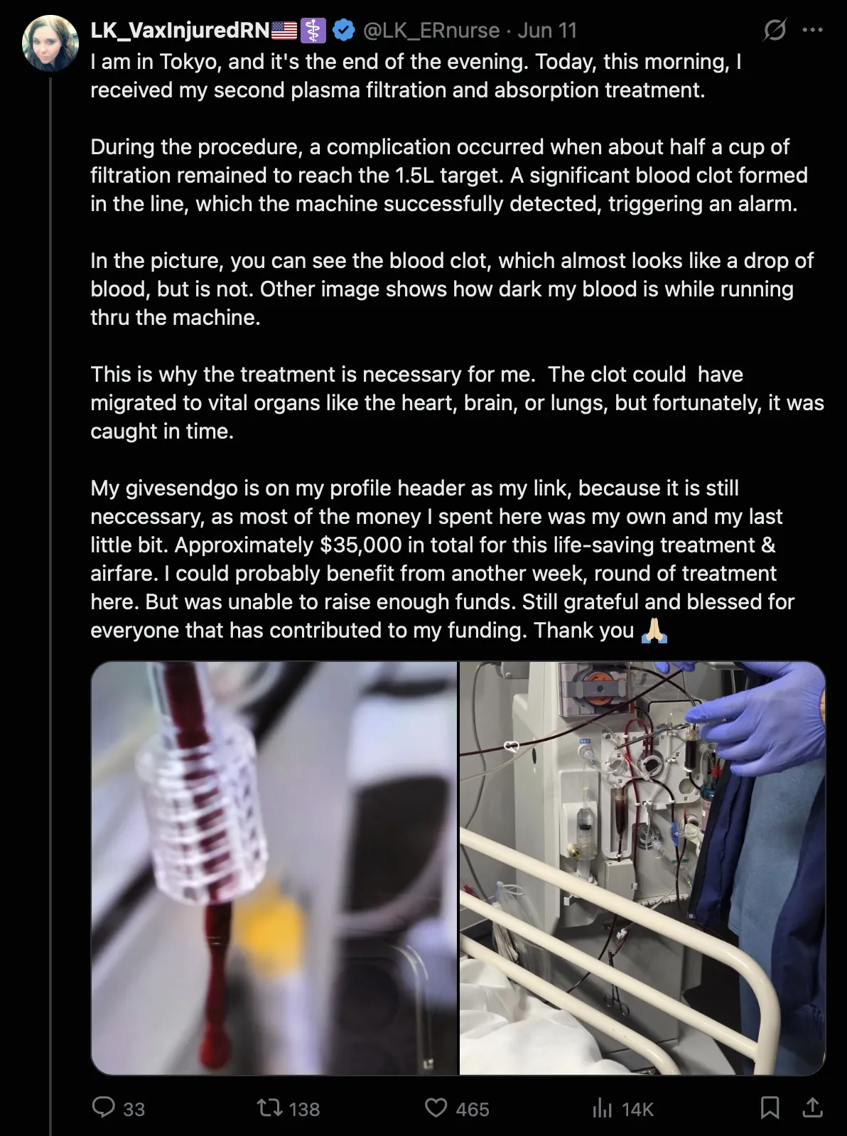
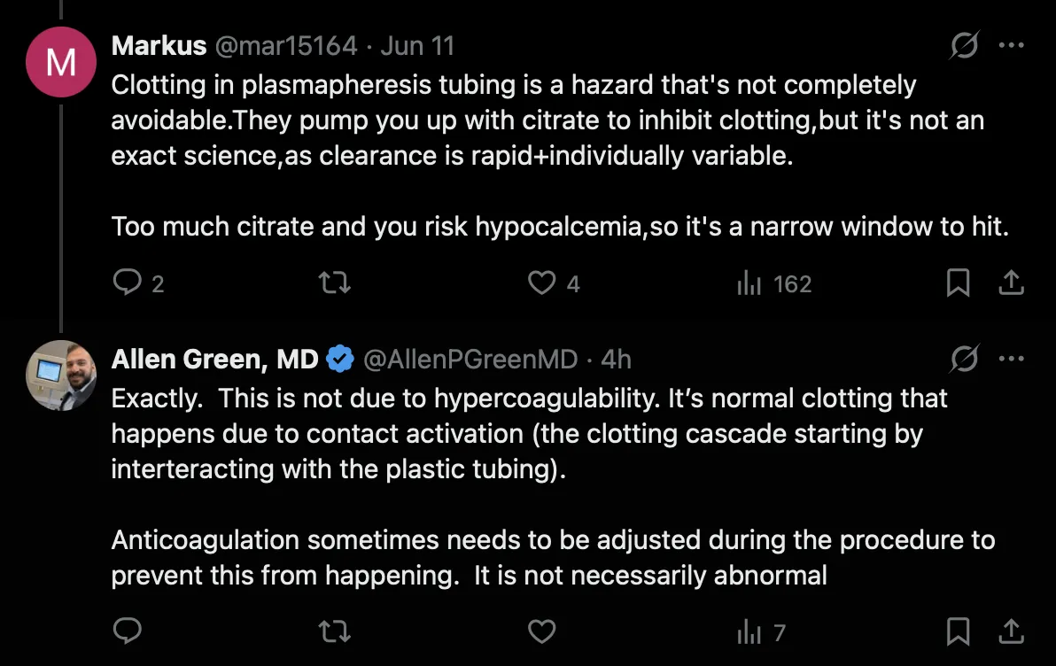
One of McCairn's patients said that while she was undergoing the treatment in Tokyo, a clot formed in the tubes of the plasmapheresis machine, and she said: "This is why the treatment is necessary for me. The clot could have migrated to vital organs like the heart, brain, or lungs, but fortunately, it was caught in time." But Markus and Allen Green pointed out that it's normal for clots to form in the tubes of a plasmapheresis circuit: [https://
One of McCairn's patients now said: "During DFPA #2 on 7/28 a small injury occurred to my right lung leading to a pneumothorax." [https://
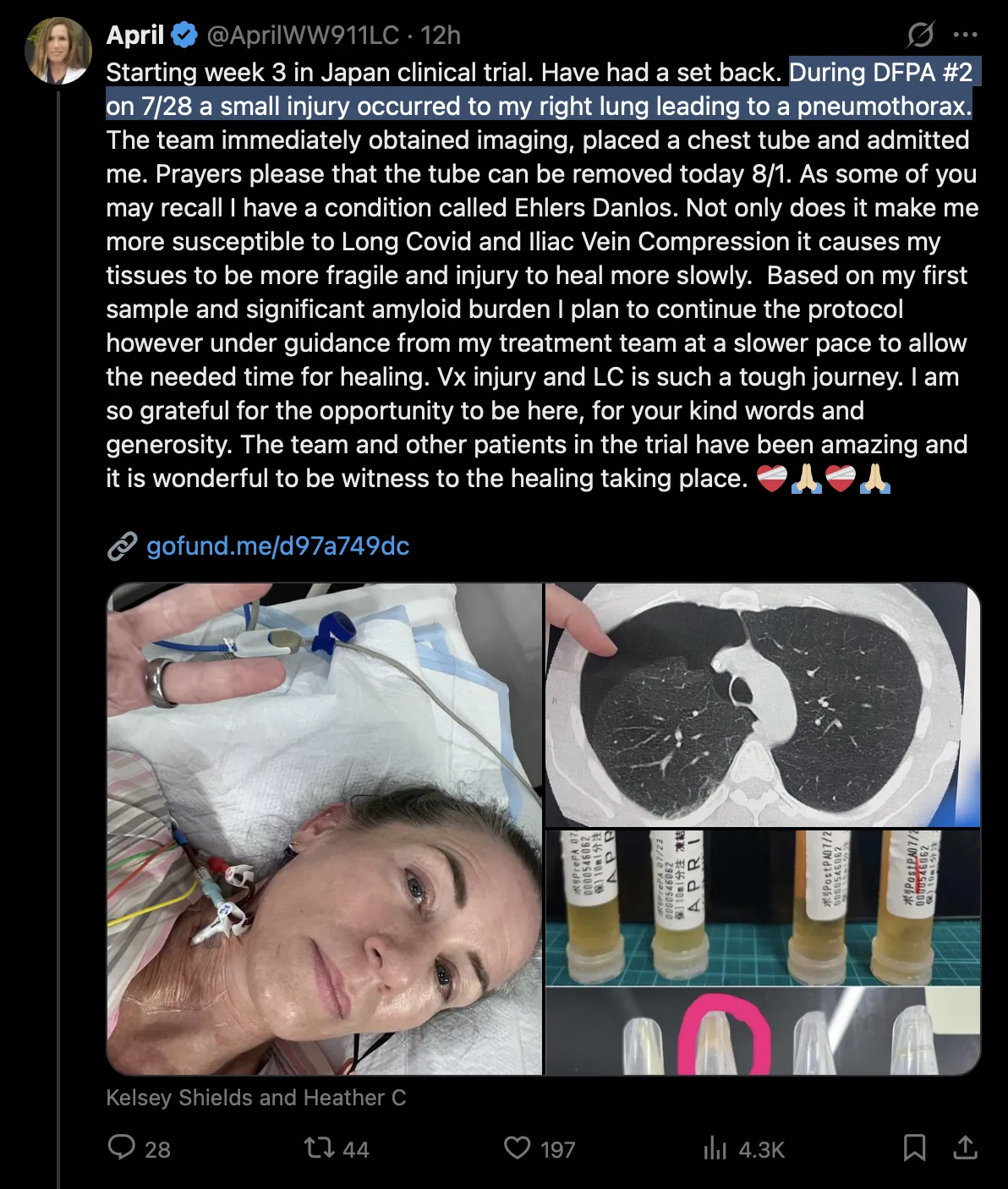
Another one of McCairn's patients was asking for donations so the patient with a collapsed lung could undergo a full 4-week treatment instead of only a 2-week treatment: [https://

When the patient whose lung collapsed during the second treatment had undergone a single treatment, she said on GoFundMe: "Many of you have asked if I'm feeling better. The honest answer is...it's still too early to know." So she doesn't seem to have experienced a miracle cure like was reported by some of McCairn's early patients. But she was still asking for donations so she could continue the treatment for the full 4 weeks: [https://

After she had undergone two treatments, she said: "I hoped for some tiny improvement but have had little and a complication. I want to continue care which is usually 4 treatments." [https://
The patient with a collapsed lung said that she was part of a "clinical trial" in Japan, so someone on Twitter was wondering why the patient was raising money if she was part of a clinical trial, because clinical trials should normally be free: [https://

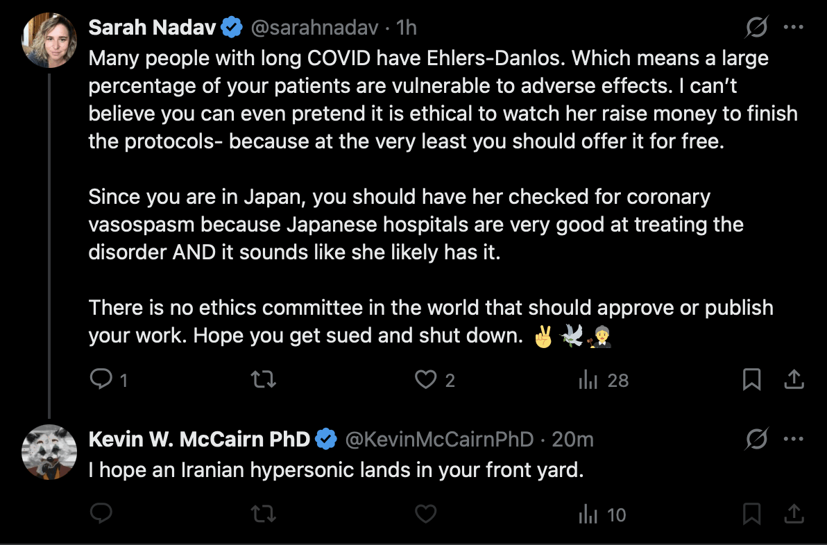
However another one of McCairn's patients responded: "Nobody is hiding the fact that it's an experimental research protocol. If someone casually used the words 'clinical trial' in a social media post, that doesn't suddenly change what it is." [https://
But anyway, McCairn has had about 50 patients so far, but at least two of them have had a severe adverse event during the treatment, because earlier Lyndsey House experienced a hypovolemic shock during the treatment. So that's a rate of around one severe adverse event per 25 patients, or possibly even more in case other patients have also experienced a severe adverse event that was not publicized.
When the Twitter user Markus told McCairn that the German IAMPOCO trial failed to find a significant benefit for immunoadsorption, McCairn said it was because the patients of the trial were not injected with the baby tooth extracts, and because not all patients had the catheter hooked up to the jugular vein.
McCairn has been saying that the reason why his plasmapheresis machine is hooked up to the jugular vein is because it somehow improves the removal of toxins from the brain. Markus told McCairn that his explanation makes no sense, and a jugular catheter is commonly used in plasmapheresis but not for the purpose of removing toxins from the brain: [https://
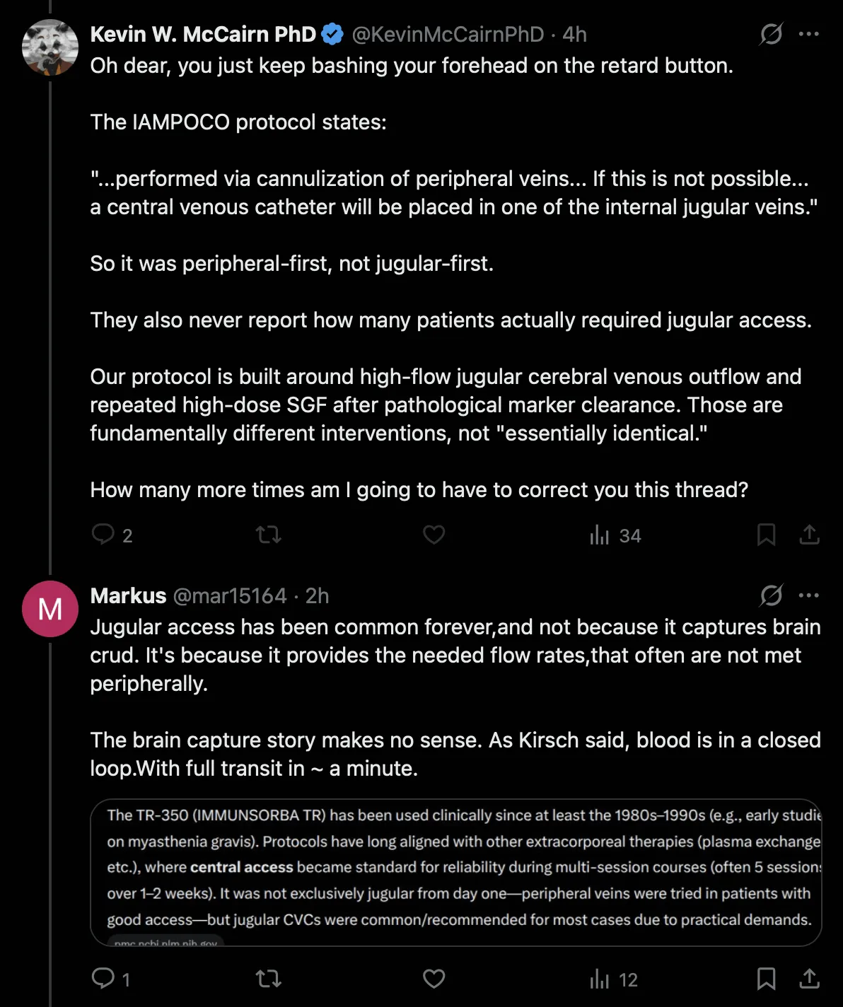
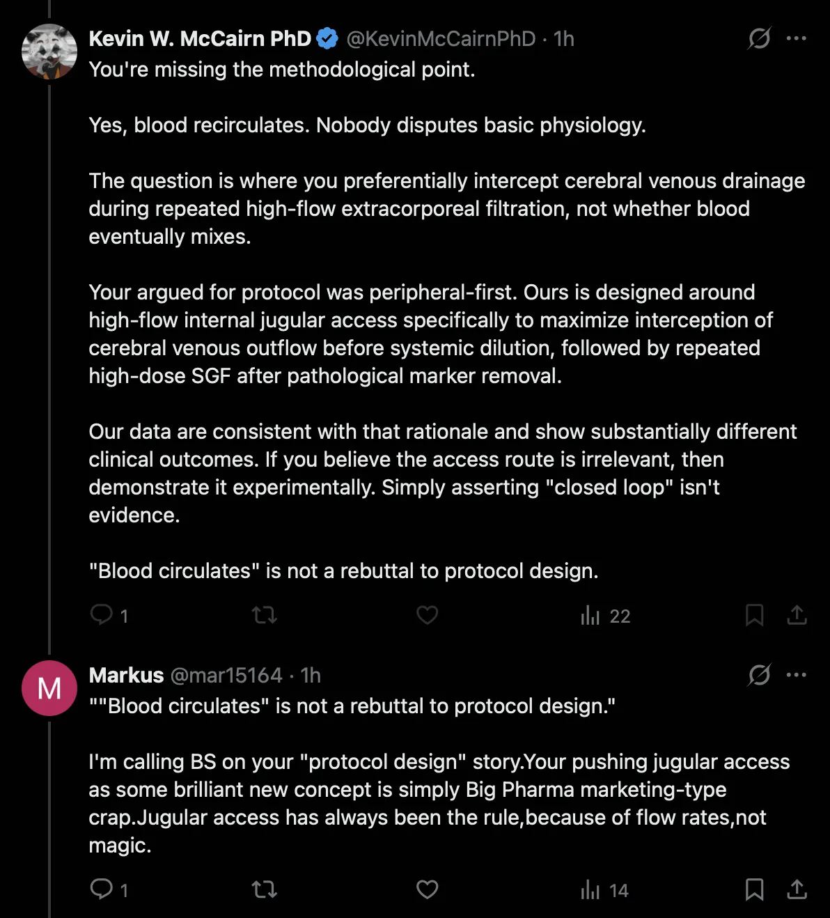
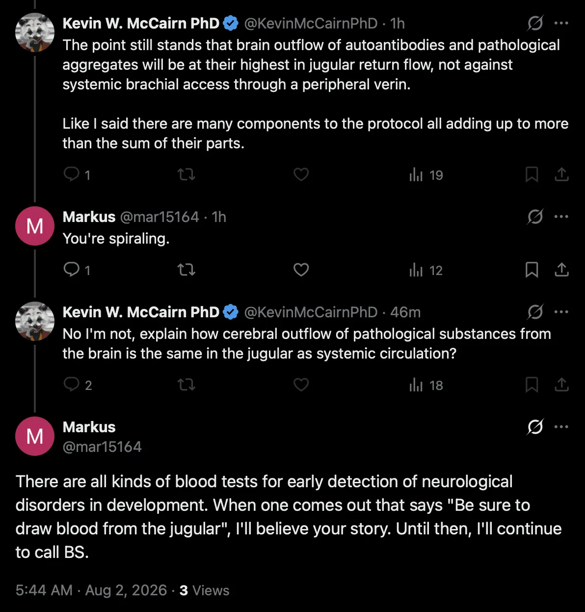

Google AI said:
An internal jugular central venous catheter is used in plasmapheresis primarily to provide the exceptionally high blood flow rates and mechanical stability required by apheresis machines.
While peripheral veins (arms) are preferred whenever possible, many patients require a jugular central line due to poor vein quality or the need for multi-day treatments.
The specific reasons a jugular catheter is selected for this procedure include:
High Flow Rates and Pressure Resistance
Prevents vein collapse: Plasmapheresis pumps pull blood out of the body at high velocities, creating intense negative pressure. Small peripheral veins would collapse, whereas the large internal jugular vein can easily handle the vacuum.
Accommodates large-bore tubing: The procedure requires stiff-bodied, dual-lumen, large-bore catheters (often called Shaldon lines or VasCaths) to draw and return blood simultaneously.
Anatomical and Safety Advantages
Straight path to the heart: The right internal jugular vein offers a direct, vertical path straight down into the superior vena cava. This minimizes the risk of the catheter kinking or hitting vessel walls, ensuring smooth blood flow.
Lower risk of stenosis: Compared to a subclavian vein catheter (in the chest), jugular catheters carry a significantly lower risk of causing long-term vein narrowing (stenosis) or thrombosis.
Lower risk of infection: Jugular lines have a lower incidence of infection and lower complication rates compared to femoral vein catheters (placed in the groin).
Patient comfort: A neck catheter allows the patient to keep their arms free and moving during multi-hour, multi-session treatments, which is impossible with large needles in both arm bends.
It's not clear if the majority or minority of patients in the German trial had the catheter connected to the internal jugular vein, even though in the study protocol that was written before the trial was conducted, a section about the sham apheresis treatment said: "In some patients, adequate blood flow can be achieved by cannulation of peripheral veins. However, in most patients, placement of a Shaldon catheter into the internal jugular vein is necessary. Shaldon catheter placement carries other potential risks such as injury to the lung resulting in pneumothorax or mispuncture of the carotid artery. However, since the catheter placement is sonography-guided, the risks for such adverse events are minimized." [https://
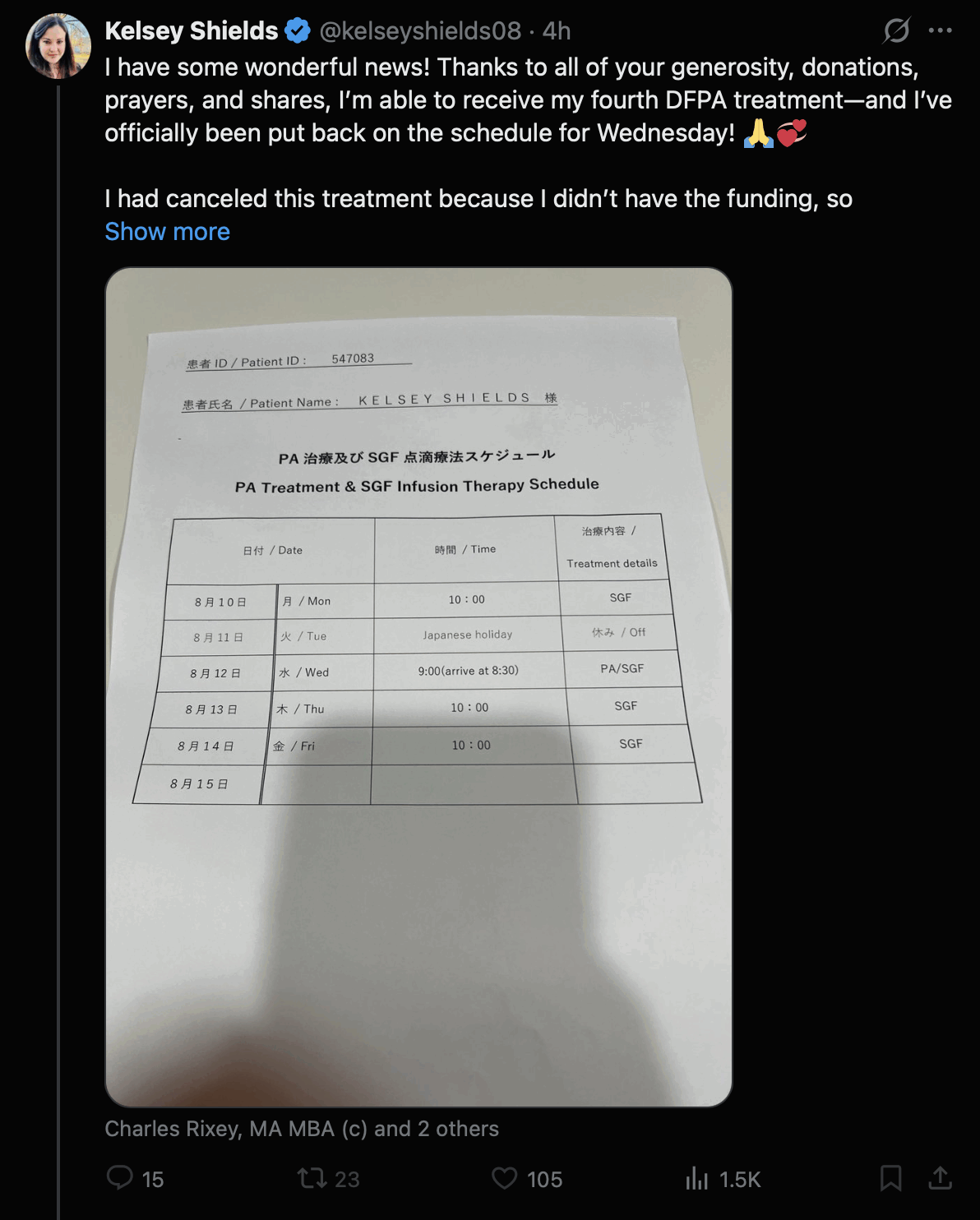
In a Japanese tweet from August 2026, the plasmapheresis treatment provided by Edogawa Hospital was now described as PA (plasma adsorption) and not DFPA (which is variously defined as either "double filtration plasma apheresis" or "double filtration plasma adsorption"). I don't know if it's because I tagged Katou in a thread on Twitter about how the nonstandard term DFPA was confusing, or if in Japan the term has been referred to as PA even earlier (even though the term PA is almost never used in English-language discussions of the Edogawa protocol): [https://

On this sheet of paper shown by one of McCairn's patients, the treatment was also called PA and not DFPA: [https://
Allen Green pointed out that unlike IA and DFPP, therapeutic plasma exchange might actually remove microclots from the blood. Then someone posted a photo of plasma in a tube and said: "You can see the amyloid hydrogel from DFPA in this picture. Why would anyone want albumin? It could be tainted with someone else's spike protein and who knows what else if someone got a dangerous shot lot. I'm unvaxxed. No way!" But Green said the albumin would not contain spike protein, and the photo did not show amyloid hydrogel: [https://

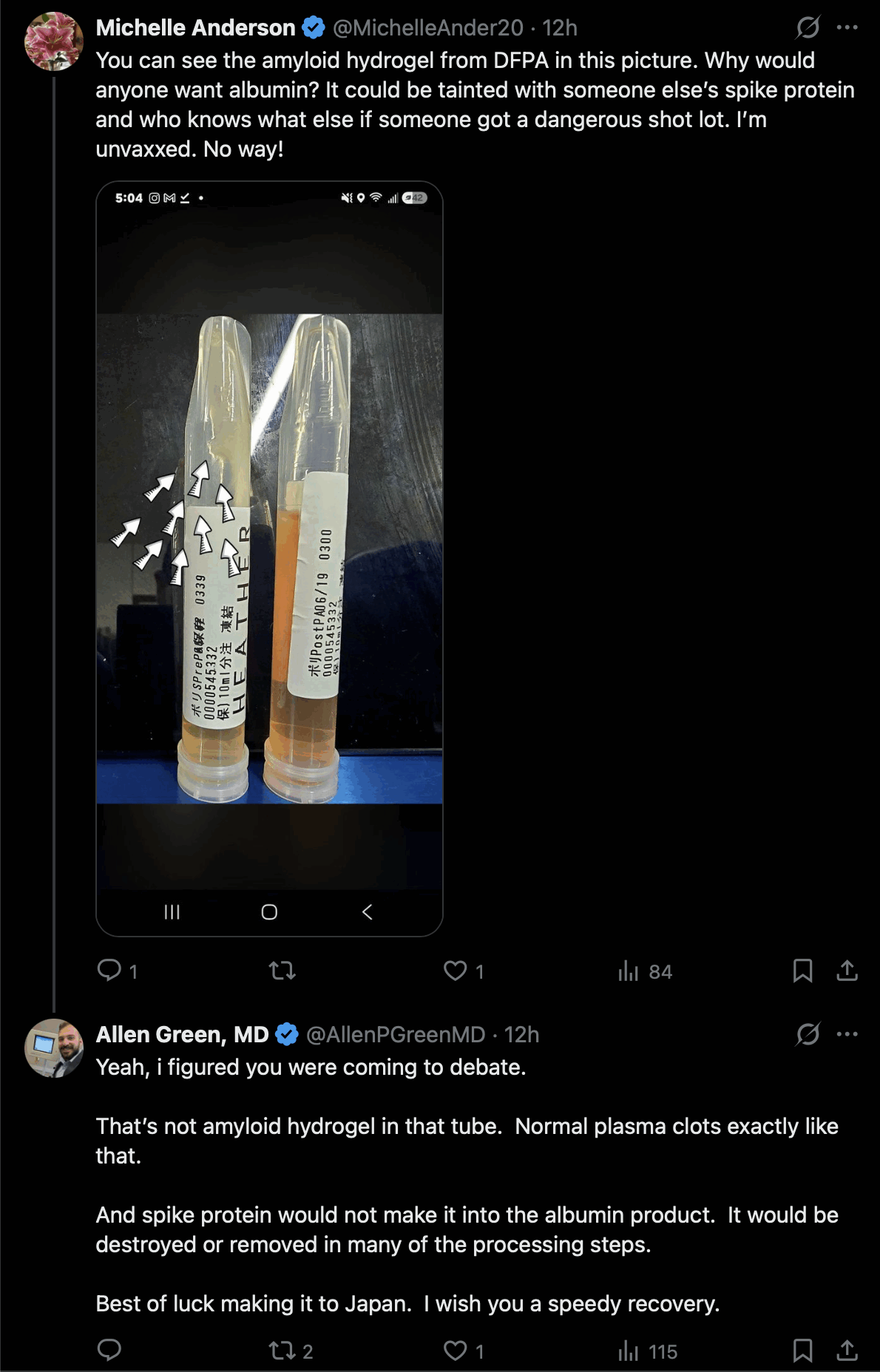
In plasmapheresis, the plasma is separated from the cellular fraction either by using a centrifuge or a membrane filter. In the case of both TPE and IA, centrifugation is more common in the United States but a membrane filter is more common in Japan.
The plasma fraction is discarded in TPE, so in TPE the centrifugation would remove the microclots if the clots remain suspended in the plasma layer, but not if the clots get spun out into the cellular layer. But it's not clear if the clots would consistently remain suspended in the plasma layer or not.
ChatGPT said that smaller fibrin microclots might be more likely to end up in the plasma layer but larger clots in the cellular layer, and that high-fibrin clots might be more likely to end up in the plasma layer but low-fibrin clots might be more likely to end up in the cellular layer:
In centrifugal TPE (e.g. Spectra Optia, COBE Spectra, COM.TEC), red blood cells (density ≈1.09-1.11 g/mL) sediment to the outside of the centrifuge, the buffy coat (white blood cells and platelets) forms the middle layer, and plasma (density ≈1.025 g/mL) remains closest to the center and is collected.
A fibrin microclot is primarily composed of protein (fibrin density around 1.3-1.4 g/mL), but it is also highly hydrated, so its effective density is only modestly greater than plasma. Its behavior during centrifugation depends largely on its size and composition. Small fibrin microclots (tens of micrometers or less) are likely to remain suspended in the plasma layer and be removed with the plasma. Larger fibrin-rich aggregates containing trapped erythrocytes or platelets are more likely to sediment toward the cellular layers and remain with the blood cells.
In membrane filtration TPE, plasma passes through membrane pores while cells are retained. Fibrin microclots that are smaller than the pore size may enter the plasma fraction, whereas larger aggregates may be retained on the membrane or contribute to filter fouling.
Most intravascular fibrin microthrombi are not composed of pure fibrin. They often contain platelets, red blood cells, leukocytes, von Willebrand factor, extracellular DNA, and other plasma proteins. These additional components increase their size and alter their sedimentation behavior.
If the aggregates are sufficiently large or contain many red blood cells, they are more likely to partition with the cellular fraction during centrifugation. Conversely, small fibrin-rich aggregates with little cellular material are more likely to remain in the plasma fraction.
Therefore, if the question is whether TPE removes circulating fibrin microclots, the expectation is that small fibrin-rich aggregates would largely be removed with the discarded plasma, whereas larger or cell-rich thrombi may partition partly into the cellular fraction and be returned to the patient.
To my knowledge, there are very few published studies that have directly measured the partitioning of fibrin microclots during centrifugal TPE, so this conclusion is based primarily on the physical principles of density and particle size rather than direct experimental evidence.
Grok said that fibrin-rich microclots would likely remain in the plasma layer, and in fact when Pretorius analyzes microclots in plasma, she uses centrifugation to separate the plasma and microclots from the cellular fraction:
In standard blood centrifugation used for therapeutic plasma exchange (TPE/plasmapheresis) or laboratory preparation of platelet-poor plasma (PPP), fibrin-rich (amyloid/fibrinaloid) microclots remain in the plasma supernatant rather than sedimenting into the cellular pellet (RBCs, WBCs, and platelets).
These microclots are routinely detected and quantified in PPP prepared by centrifugation (commonly ~3,000 x g for ~15 minutes). Researchers such as Pretorius, Kell, and others isolate PPP this way and then visualize the microclots (via thioflavin T or similar stains, fluorescence microscopy, or imaging flow cytometry). Higher-force centrifugation of the plasma itself (e.g., ~20,000 x g) can later pellet them for proteomic analysis, confirming they stay suspended under the milder forces that separate cells from plasma. [https://
pmc. ]ncbi. nlm. nih. gov/ articles/ PMC11131067/ They are typically 1-200 um in size (median often around ~15 um). Although this overlaps with cellular dimensions, their density, structure, and composition mean they do not reliably co-sediment with the cellular fraction under the conditions used for plasma separation in TPE or PPP preparation. [https://
www. ] In TPE, the separated plasma (containing soluble proteins, inflammatory mediators, autoantibodies, fibrinogen-related factors, and these microclots) is removed and replaced with albumin or donor plasma, while the cellular components are returned to the patient. This is one reason TPE has been explored experimentally for conditions associated with circulating microclots (e.g., Long COVID research contexts).frontiersin. org/ journals/ bioengineering- and- biotechnology/ articles/ 10. 3389/ fbioe. 2025. 1604447/ full
Allen Green said: "The median microclot is supposed to be around 20 microns. I'd suspect many at that size would remain in the plasma during centrifugation, but would want to see literature to confirm." [https://
Allen Green wrote the following in his blog: [https://
We know that plasma contains pro-aging, pro-inflammatory factors that accumulate over time and contribute to biological aging. Some of these have been identified: certain inflammatory cytokines, immune complexes, dysfunctional proteins. But the full catalog is unknown. We know these factors exist and that they matter not because we have mapped all of them, but because when we remove plasma broadly and replace it, without knowing exactly what we removed, tissues rejuvenate.
The animal evidence for this is striking. Heterochronic parabiosis experiments, in which the circulatory systems of old and young mice are surgically joined, demonstrated as early as 2005 that old tissue can be rejuvenated by exposure to a younger blood environment. For years the assumption was that young blood contained beneficial factors. But a landmark 2020 study published in Aging (Mehdipour et al.) overturned that interpretation. The researchers performed neutral blood exchanges on old mice, replacing their plasma not with young blood but simply with saline and albumin. A single exchange rejuvenated muscle, brain, and liver, tissues derived from all three embryonic germ layers. Pro-aging protein signatures decreased, pro-regenerative ones increased, and the benefits were comparable to what parabiosis had achieved. The implication was clear: you do not need to add anything from young blood. You need to remove what has accumulated in old blood. This is, in effect, the animal model of therapeutic plasma exchange.
The clinical translation followed. Dr. Kiprov's group demonstrated in the 2022 GeroScience study (Kim et al.) that old plasma dilution reduced measurable biological age in human patients. The 2025 Aging Cell study (Fuentealba et al.), also from Dr. Kiprov's research program, extended this finding using 35 epigenetic clocks, showing that TPE combined with IVIG produced an average reduction in biological age of approximately 2.61 years. These benefits emerged from broad, non-selective removal, not from targeting specific known molecules.
Dobri Kiprov is the director of the apheresis center where Green works. These were the two studies by Kiprov's group Green referred to: https://
I believe Beate Jaeger was the pioneer of trying to use plasmapheresis to remove the fibrinaloid clots described by Pretorius and Kell. But Jaeger's HELP apheresis treatment seems to use a membrane filter and not a centrifuge.
One of her papers says "During HELP apheresis, blood cells are first separated from plasma in the extracorporeal circuit", and the paper says that she usesa HELP system by the manufacturer B. Braun. [https://
B. Braun sells only a single HELP system called Plasmat Futura, which uses a plasma filter called Haemoselect that has a pore size of 0.5 to 0.7 µm. B. Braun's website says: "The Haemoselect 0.5 / 0.7 plasma filter with a hollow fiber consisting of polyethersulfon is an integral part of the HELP apheresis treatment unit, and is used for plasma separation in HELP apheresis on the Plasmat Futura machine." [https://
Grok said:
Beate Jaeger's HELP apheresis treatments rely on the commercial B. Braun Plasmat Futura system. In this setup, whole blood first passes through the Haemoselect membrane plasma filter, which separates plasma from the cellular components before any further processing occurs. Manufacturer specifications list the pore size of this Haemoselect filter as 0.5 to 0.7 micrometres. The separated plasma is then mixed with high-dose heparin and acetate buffer to lower its pH, causing fibrinogen, LDL, VLDL and related complexes to precipitate; those solid precipitates are subsequently trapped in a downstream precipitation filter, which is the source of the visible white or yellow gel-like material observed after treatment.
Fibrin-rich amyloid microclots reported in Long COVID research typically measure between 1 and 200 micrometres, with a median size often around 15 micrometres. Because these dimensions are substantially larger than the 0.5-0.7 micrometre pores of the Haemoselect membrane, the intact microclots cannot cross into the plasma stream. Instead they remain on the cellular side of the filter and are returned to the patient together with the red cells, white cells and platelets.
Grok said:
No published quantitative before-and-after comparison of microclot burden from the Jaeger / Pretorius collaboration (or closely related group papers) has been identified.
Jaeger's main case series on HELP for Long COVID (the 2023 Gavin Publishers paper and related 2022 theoretical paper) reports:
- Clinical symptom improvement in most patients
- Reductions in fibrinogen and certain coagulation factors measured before and after sessions
However, these publications do not include systematic pre- and post-treatment quantification of microclot load using the methods Pretorius's group typically employs (thioflavin T / fluorescence microscopy, clot scoring/grading, imaging flow cytometry, or similar).
Pretorius's team collaborated with Jaeger starting around late 2021 specifically to evaluate whether HELP could address the microclots they had described, and they developed grading systems for microclots and platelets. Despite this, no peer-reviewed paper from the group presenting paired before-and-after microclot burden data after HELP treatment appears to have been published.
One of Beate Jaeger's papers said: "Additionally, HELP apheresis has been found to remove circulating fibrinolysis-resistant microclots [18,19]." [https://
When I asked if other groups had measured microclot burden before and after apheresis treatment, Grok said:
Here are sources for the later clinic or independent reports that claim reductions in microclot-related measures after apheresis (distinct from the original Jaeger-Pretorius collaborative publications):
1. Dr Yannick Kok / FibriScore presentation (reported by Philip McMillan)
https://
drphilipmcmillan. substack. com/ p/ lessons- from- the- front- line- apheresis This conference summary states that Dr Yannick Kok presented laboratory evidence that microclot complexes, measured by his FibriScore® test, "drop significantly after apheresis."
2. Apheresis Center (Cyprus) claims
https://
apheresiscenter. eu/ blog/ help- apheresis- paper- removes- spike- protein- microclots- more The clinic states that research microscopy shows platelet hyperactivation and microclot-like / amyloid-like structures "dwindle post-treatment" with HELP apheresis.
Both sources Grok linked were marketing-type materials which didn't necessarily seem credible.
The first source was a presentation by Yannick Kok, who did not actually show that the level of clots would "drop significantly after apheresis", because he only discussed the results of two patients, so the results were not significant in the statistical sense. And compared to the baseline level of the patient before treatment, the level of clots was higher a day after the treatment but lower six days after treatment, so whether the level of clots was higher or lower after the treatment depends on how many days after the treatment you're looking at.
The second source was the website of the Cyprus Apheresis Center, but the website didn't include any numeric data that would've shown if the treatment reduces the level of microclots. The center in Cyprus provides three forms of apheresis treatment, which are HELP apheresis, DFPP using the German Inuspheresis system, and immunoadsorption. But the center appears to use a membrane filter and not a centrifuge with all three methods. [https://
Yannick Kok works for Ayus Laboratories, which is the German company that manufactures the Inuspheresis DFPP system. But Inuspheresis would likely not remove microclots, because it employs a membrane filter and not a centrifuge so separate the plasma. [https://
Ayus Laboratories has developed a test called FibriScore to quantify microclot level. Philip McMillan wrote: [https://
Ayus Laboratories' FibriScore® test uses imaging flow cytometry with fluorescent dyes that bind to amyloid structures, quantifying both the number and size of FMCs in a blood sample. For the first time, clinicians can see what's actually flowing through the pipes rather than inferring from indirect measurements.
The Cytek ImageStream® technology combines the statistical power of flow cytometry with the visual clarity of microscopy. Every particle passing through the machine is photographed, measured, and characterized. The result: patient reports show dramatic differences between healthy individuals (minimal FMCs) and long COVID patients (sometimes three-fold elevations in total microclot area).
FibriScore® is particularly valuable for treatment monitoring, revealing whether interventions are working. Post-apheresis samples show marked reductions, visible proof that the cleaning worked. One patient Kok tracked showed total microclot area dropping from 49.30 mm² to 7.60 mm² six days after treatment. Another showed sustained improvement over five months, from 60.09 mm² to 20.31 mm².
It's weird how McMillan only wrote about the results of two individual patients, and not multiple patients averaged together. The 6-fold variation of the first patient and the 3-fold variation of the other patient might be within the normal range of day-to-day variation in clot level, so the difference was not necessarily because of the treatment.
During an interview with Philip McMillan in 2025, Yannick Kok showed the image below, and he said: "I'm part of the Ayus Medical Group, and the thing of the Ayus Medical Group is that we do apheresis, and specifically we do Inuspheresis. And a big question for us is, is Inuspheresis able to filter out these microclots from a patient? I've used the TKM-58 filter in the Inuspheresis, and the filter - the pore size is in the nanometer range in this filter, and microclots they are in a micrometer range. So from a physical, uh, if you think of the physics, big particle, small hole, doesn't fit. So what [unintelligible] we would expect. We hook up a patient with high microclots, um, and after aphoresis we should not detect these high microclots anymore. Then we got this data. And this was a surprise. So here in gray, you see the initial value before aphoresis. In red, you see the value directly after Inuspheresis, and we see an increase. And we got worried. We were surprised to see this. And we started to think, how can this be? Because if you think of the physics behind it, we should remove it." [https://
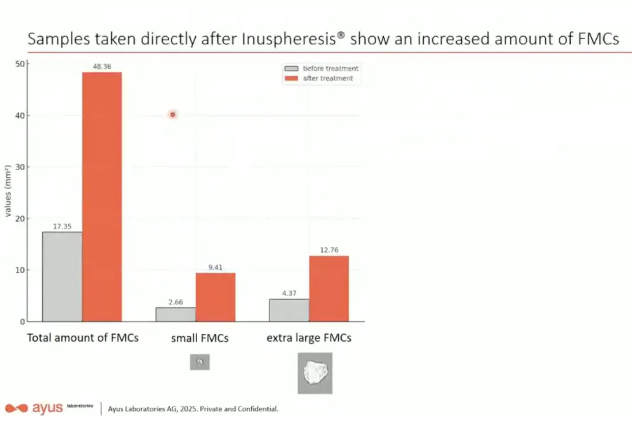
Did he really not understand the basic mechanics of his own company's product? The TKM58 filter is the plasma fractionator filter, which is the second filter employed in DFPP. [https://
When Yannick Kok said that the patient had a higher microclot level a day after treatment than before treatment, he was talking about data for a single patient only, so the increase may have been due to random day-to-day variation in the microclot level.
But anyway, next Kok showed that when he measured the microclot level 6 days after treatment, the level was about 6 times lower than before treatment. And he said it was consistent with how a patient who has undergone the treatment will not necessarily feel better immediately after the treatment, but only a few days later.
But he again only showed data for a single patient and not multiple patients averaged together, so even the level after 6 days might have been within the normal range of day-to-day variation.
So basically his sample size was too small to draw conclusions about how Inuspheresis affects the microclot level, even though if you understand that the clots are too big to pass through the plasma separator, then it makes sense why the level of the clots was not reduced a day after the treatment.
Added later: I now found a paper by Beate Jaeger from 2023, which included measurements of the level of microclot-like structures before and after HELP apheresis in 16 subjects. There was no significant reduction in the area of the microclot-like structures (p-value about 0.25) but only in the count of the microclot-like structures (p-value about 0.01): [https://
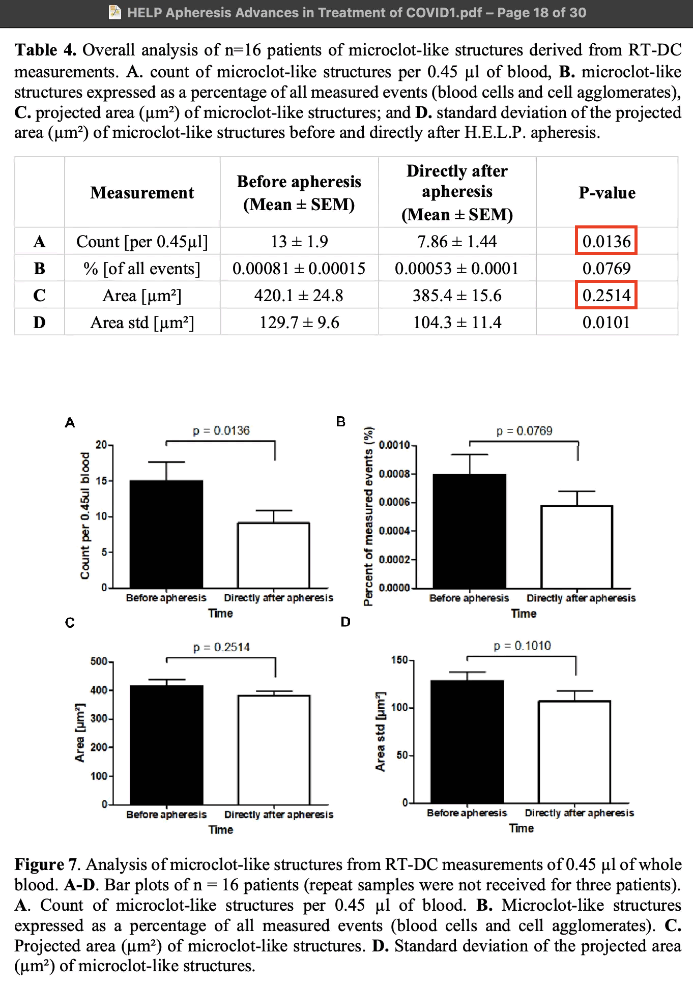
The so-called microclot-like structures were identified using a method of cytometry called real-time deformability cytometry (RT-DC), so that the authors selected structures with an area at least 100 µm² and a low deformation parameter relative to the area. The deformation parameter indicates how much a structure changed in shape as it moved through the measurement channel. But the method of identifying microclots may have been prone to false positives, because the authors wrote: "it should be kept in mind that not all events are necessarily clots, as damaged endothelial cells or cellular debris generated by the apheresis process may have been present and therefore recognized in the analysis". The so-called microclot-like structures were not selected based on whether they were fluorescent or stained by Thioflavin. And since only structures with an area above 100 µm² were classified as microclot-like structures, all of the structures would've likely been too big to pass through the pores of Jaeger's plasma separator, which likely has a pore size of 0.5 to 0.7 µm: [https://
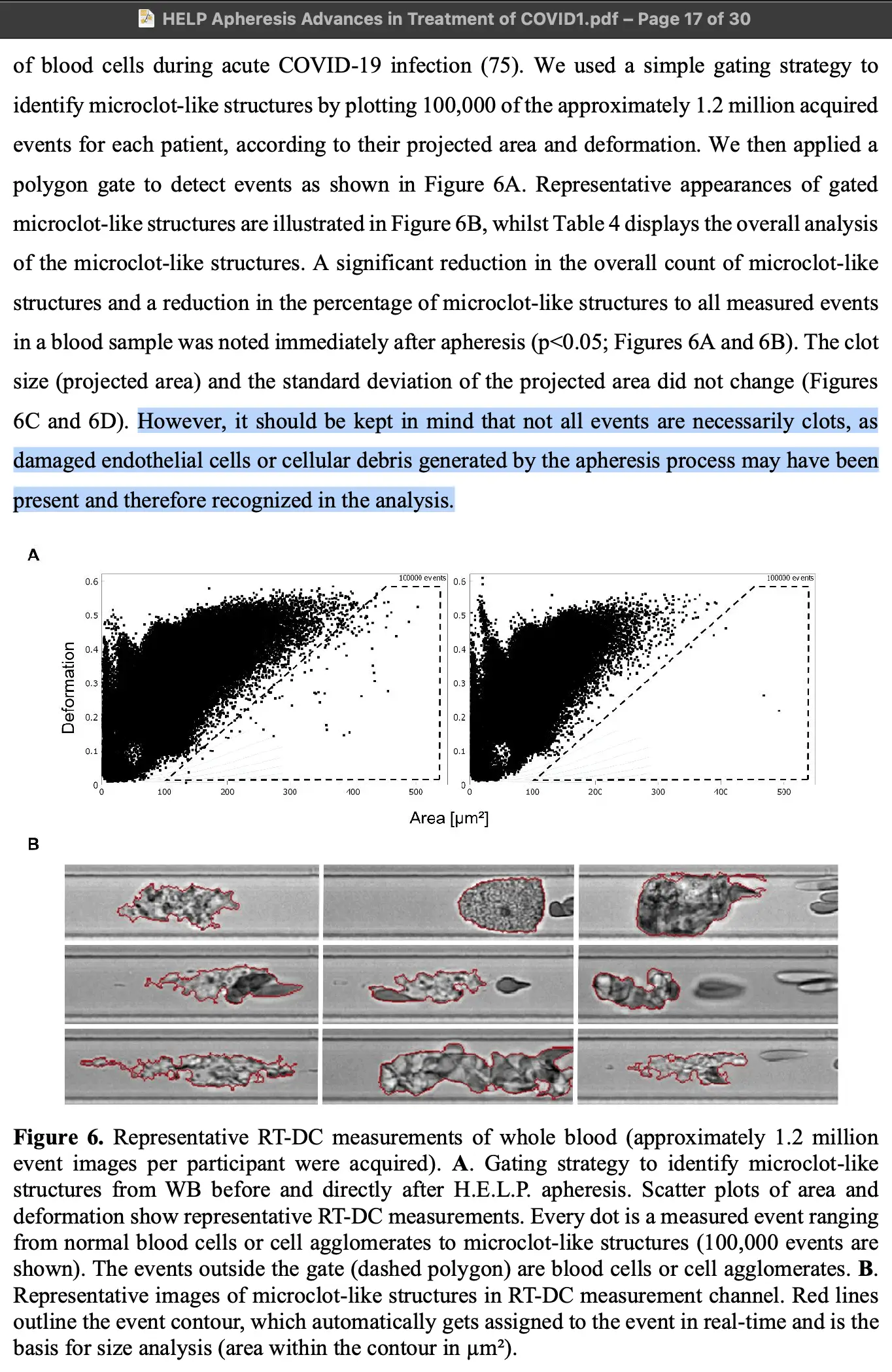
McCairn claims that the IA treatment provided at Edogawa is somehow safer than the TPE treatment provided in the United States, because in TPE the patient is given replacement albumin, and he claims the albumin is contaminated with spike protein or microclots. However I have never seen him present evidence that albumin products contain spike protein of SARS-CoV-2.
Here when he claimed that albumin had been found to contain spike protein, Allen Green asked: "Albumin from which manufacturer? Mind sharing the reports?" But McCairn never answered: [https://

One of McCairn's followers said that albumin has been found to bind spike protein in some laboratory studies. But Allen Green pointed out it doesn't mean that the albumin sold by manufacturers would contain spike protein: [https://

Then McCairn's follower said that blood banking protocols don't screen for the presence of spike protein, but Allen Green pointed out that the screening is not necessary because the spike protein is removed by the manufacturing process:
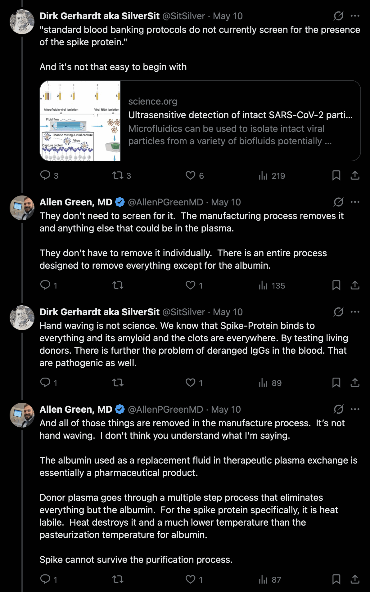
Then McCairn claimed that every batch of blood bank products he had tested had been positive for "cross-seeding amyloidogenic fragments". When Allen Green asked to see the evidence, McCairn didn't show the evidence but he said "I do all the time. You should pay closer attention to my output." However I have paid close attention to McCairn's output, and I don't remember seeing him post the evidence:
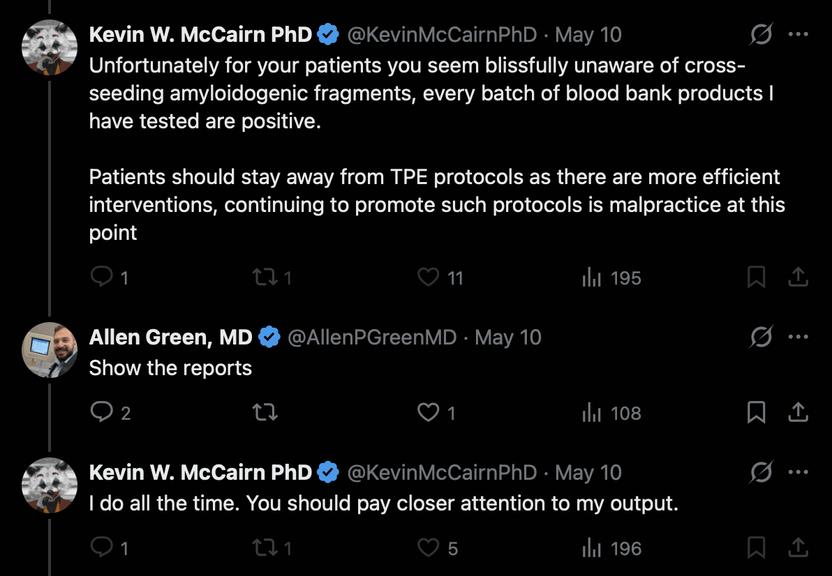
Google AI said:
No, the SARS-CoV-2 spike protein does not survive the rigorous manufacturing and purification processes required to produce human serum albumin. The spike protein, like the virus itself, is highly vulnerable to the harsh physical and chemical steps designed specifically to eliminate viral and protein contamination during plasma fractionation.
Human serum albumin is manufactured using the Cohn cold ethanol fractionation method or advanced chromatographic purification, both of which utilize concentrated alcohol and extreme pH adjustments to selectively precipitate and separate proteins. These chemical conditions disrupt the structural integrity of the spike protein, which is a fragile trimeric glycoprotein. Furthermore, the purification process explicitly includes a robust viral inactivation phase called pasteurization, where the albumin solution is heated to exactly 60 °C (333.15 K) for a continuous period of 10 hours.
While native albumin is uniquely heat-stable and survives this thermal stress with the aid of stabilizers like caprylic acid, the SARS-CoV-2 spike protein denatures rapidly at much lower temperatures. This pasteurization step completely unfolds the secondary and tertiary structures of any residual spike protein, rendering it non-functional and completely degraded. As a result, the final therapeutic albumin product has an exceptional safety profile, completely free of intact or active SARS-CoV-2 viral components.
Google AI also said: "No scientific study has ever found the SARS-CoV-2 spike protein in commercially manufactured albumin products. Global regulatory bodies and researchers have continuously monitored plasma-derived therapies throughout the pandemic, and zero instances of spike protein contamination have been reported in finished therapeutic human serum albumin."
Earlier when McCairn has been asked why the catheter is hooked up to the jugular vein in the Edogawa protocol, he has given the implausible-sounding explanation that the jugular catheter somehow helps to capture toxic substances that are released from the brain.
Now he came up with another similarly implausible explanation, because McCairn said that the jugular catheter would somehow enhance a mechanical pumping action that causes fibrin deposits to be released from muscle tissue into blood vessels: [https://
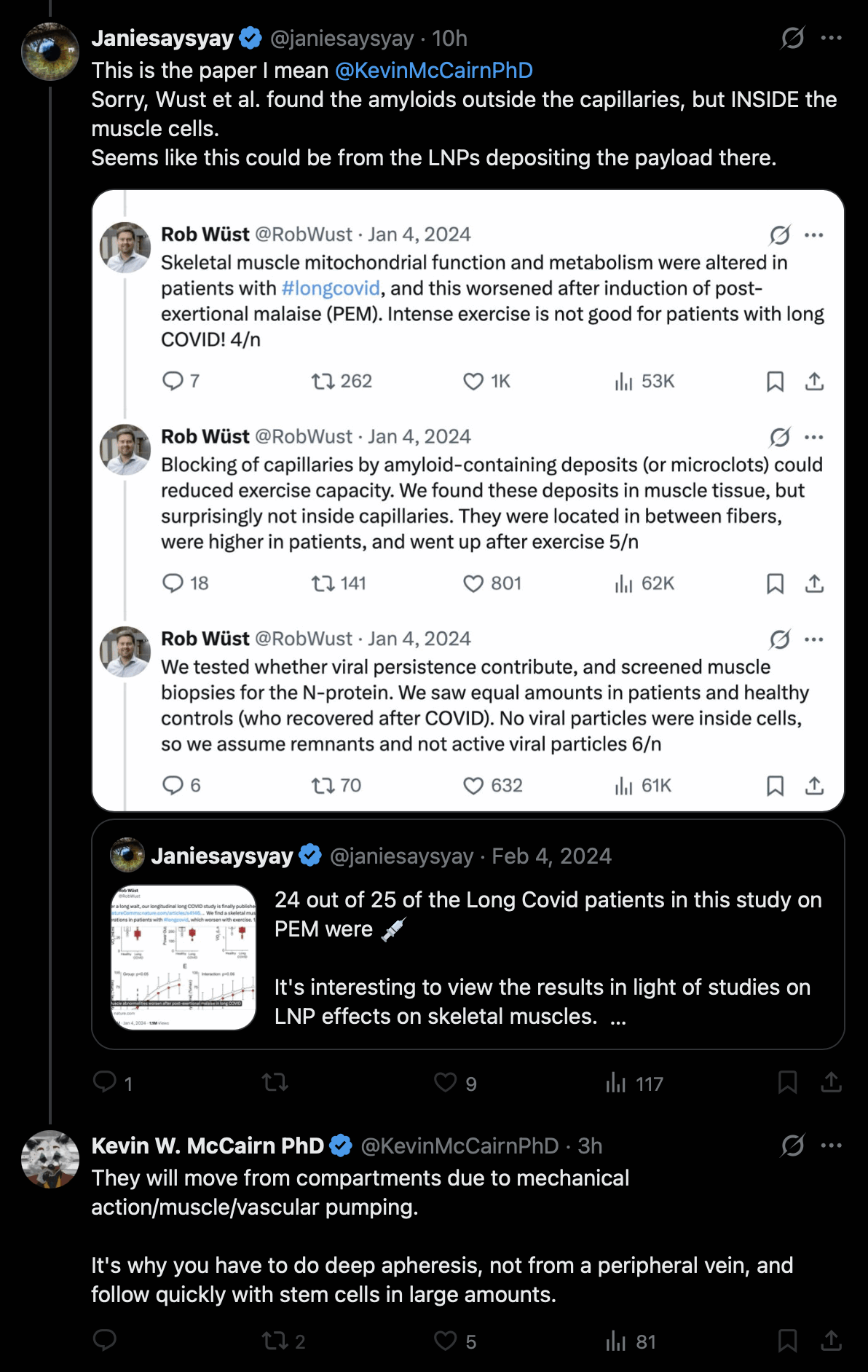
When I asked if his explanation was plausible, Grok said:
Fibrinaloid (amyloid-containing) deposits located in the interstitial space of skeletal muscle would not be expected to migrate into blood vessels as a direct result of the mechanical forces of plasmapheresis performed via a jugular catheter.
What the relevant study actually found
In the 2024 Nature Communications paper by Appelman, Wüst and colleagues, amyloid-containing deposits were present at higher levels in the skeletal muscle of long COVID patients than in recovered controls. These deposits sat between muscle fibers (extracellular/interstitial), not inside capillaries, and their density increased after an exercise challenge that induced post-exertional malaise (PEM). The authors explicitly noted they were not located inside capillaries and therefore were not observed to be directly occluding them in the biopsy samples. [https://
www. ]nature. com/ articles/ s41467- 023- 44432- 3 Why the proposed migration mechanism does not hold
Location matters. Interstitial deposits sit outside the vascular compartment, separated from circulating blood by the capillary endothelial barrier and basement membrane. Large, insoluble, amyloid-like fibrin aggregates do not freely diffuse across intact endothelium.
Apheresis mechanics are intravascular. Therapeutic plasma exchange (or double-filtration plasmapheresis) withdraws blood from a vein, separates plasma, removes selected components, and returns the remainder. Whether access is peripheral or central (internal jugular), the flow and shear forces act only on the blood already inside the vascular system. The procedure does not create pressure gradients, "pumping," or mechanical agitation capable of forcing distant interstitial material from muscle tissue into capillaries or larger vessels.
Central (jugular) access is chosen for practical reasons, not tissue mobilization. High-flow central catheters support the blood-processing volumes needed for effective treatment when peripheral veins cannot deliver adequate flow or are otherwise unsuitable. This is a standard technical consideration in apheresis, unrelated to extracting tissue-bound deposits.
Physiological mobilization is a separate issue. Muscle contraction, local inflammation, or changes in vascular permeability could theoretically allow some soluble factors or small particles to enter lymphatics or blood over time. That process, if it occurs at all for these deposits, would be driven by ordinary physiology or pathology - not by the presence or mechanics of a jugular catheter during apheresis.
In short, plasmapheresis can only remove what is already circulating in the blood. Claims that the mechanical action of "deep" (central-access) apheresis specifically mobilizes interstitial muscle fibrinaloid deposits into the vasculature lack support from vascular physiology, apheresis practice, or the muscle-biopsy findings themselves.
In the Twitter thread about the paper, the senior author Wüst referred to the so-called deposits as "microclots", because he wrote: "Blocking of capillaries by amyloid-containing deposits (or microclots) could reduced exercise capacity. We found these deposits in muscle tissue, but surprisingly not inside capillaries." [https://
Grok said:
Wüst and colleagues did not show that the amyloid-containing deposits they observed in muscle were fibrin (or fibrinaloid microclots).
What they actually did
In the 2024 Nature Communications paper (Appelman et al.), the team detected the deposits using Thioflavin T (ThT) staining on skeletal muscle biopsy sections. Thioflavin T is a fluorescent dye that binds to β-sheet-rich structures and is a general marker for amyloid material of various protein origins. They also co-stained some sections with antibodies against CD31 (to mark endothelial cells/capillaries) and LYVE-1 (to mark lymphatic vessels) to determine the location of the deposits.
They consistently described the structures only as "amyloid-containing deposits." They reported that these were more abundant in long COVID patients' muscle (extracellular matrix between fibers, next to but not inside capillaries), and that the amount increased after exercise-induced post-exertional malaise.
What they did not do
No fibrin- or fibrinogen-specific immunohistochemistry, immunofluorescence, or other biochemical identification (e.g., antibodies against fibrin(ogen), mass spectrometry of the deposits, or other fibrin markers).
No claim that the deposits were fibrin, amyloid fibrin, or the "fibrinaloid microclots" described in the Pretorius/Kell literature.
The paper references the circulating microclot hypothesis as background context but concludes that the deposits they saw were not located inside vessels and therefore did not support the idea of capillary blockage by such material. The molecular identity of the muscle deposits was left open; the authors noted that the reason for their intramuscular accumulation remained elusive.
When I asked if Wüst's group had determined in other studies if the deposits were made of fibrin, Grok said:
No. There is no published evidence that Rob Wüst's group (including collaborators such as Appelman or Charlton) has determined that the amyloid-containing deposits observed in skeletal muscle are fibrin (or fibrinaloid microclots).
Original 2024 study
In the Nature Communications paper, the deposits were identified solely by Thioflavin T staining (a general amyloid dye). No fibrin- or fibrinogen-specific antibodies, proteomics, or other compositional analyses were performed. The structures were described only as "amyloid-containing deposits."
Later work by the group
Subsequent papers and preprints from the same team focus on related topics such as: - Muscle fiber-type shifts - Mitochondrial function - Comparisons between long COVID, ME/CFS, and bed-rest deconditioning
These continue to refer to the structures as "amyloid-containing deposits" (or cite the original finding) without new data identifying their protein composition as fibrin.
Wüst has received funding (e.g., from ME Research UK) for projects exploring microclots and microvascular pathology in muscle in the context of long COVID/ME/CFS and PEM. However, no peer-reviewed publications from the group have yet reported specific identification of the muscle deposits as fibrin.
External commentary
Other researchers (particularly in the Pretorius/Kell circle) have noted that the muscle deposits "resemble" or may be related to circulating fibrinaloid microclots, but those interpretations come from outside Wüst's team and are not findings demonstrated by his group.
Some of McCairn's followers seem to think that the deposits described by Wüst had somehow formed in the circulatory system but then migrated to the muscle tissue. But I didn't find that hypothesis actually proposed by Wüst's group anywhere, and his group seems to have not answered the questions of how the deposits are formed or what the deposits are made of.
The authors appeared to assume that the deposits consisted of amyloid protein because the deposits were stained by ThT, but the authors didn't describe doing confirmatory tests like staining with Congo red or an Amytracker dye, or doing electron microscopy to see if the deposits contain amyloid fibrils.
Someone on Twitter told me I was wrong about the Edogawa protocol, because he said there were two studies that showed IA produced an improvement in the symptoms long COVID patients: [https://

But his first link was for a trial protocol for an RCT called IA-PACS-CFS, which had no results reported. [https://
The Twitter user Markus posted a video where the authors of the unpublished RCT presented their preliminary results. The primary outcome measured in the study was the change in the Chalder Fatique Scale between the baseline and approximately 60 days after treatment, but there was no significant difference in the measure between the treatment and sham groups: [https://
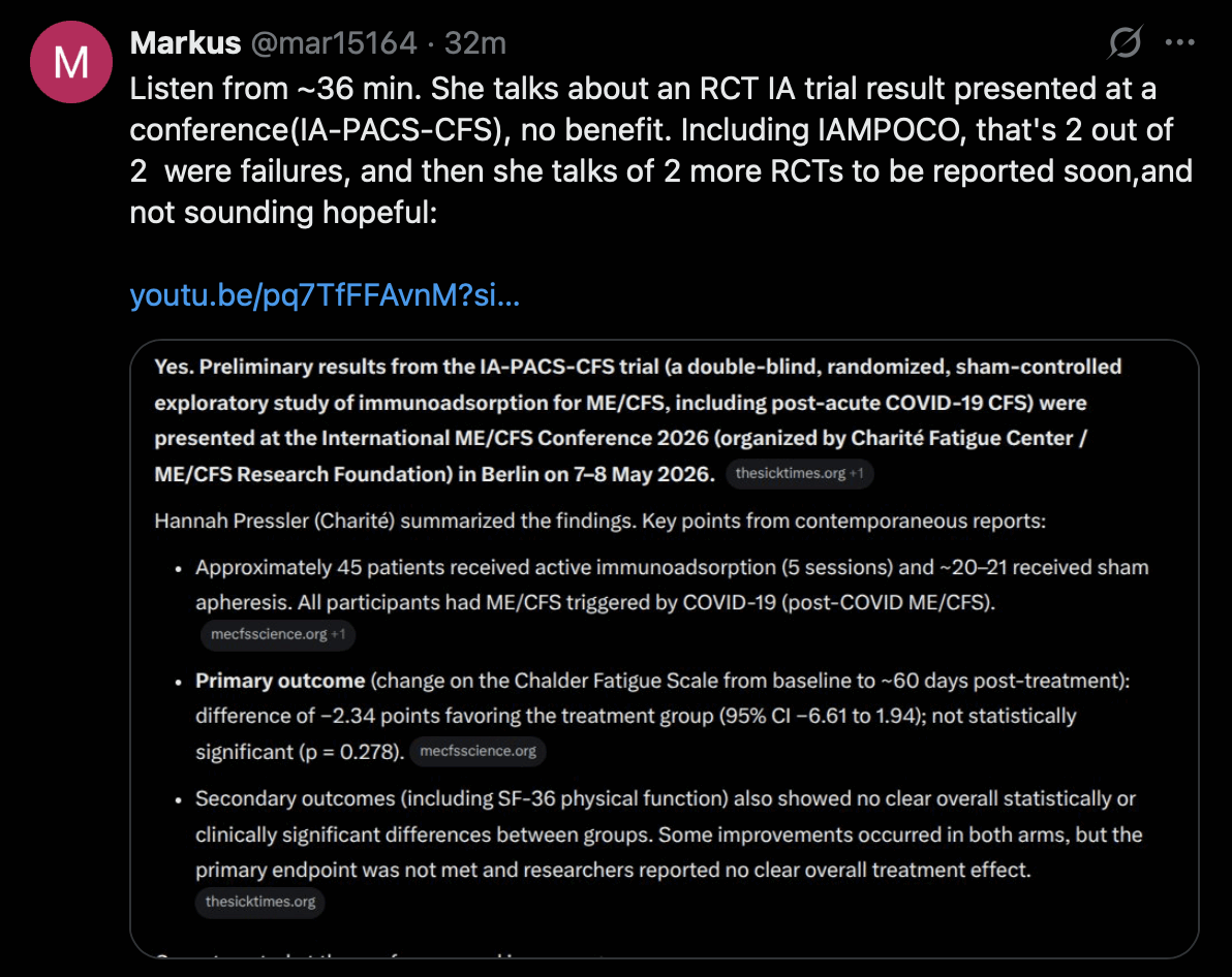
Markus pointed out how the unpublished RCT used a jugular catheter to ensure a high flow rate of blood. McCairn has been making it seem like the reason why his protocol uses a jugular catheter is because it somehow enhances the removal of harmful substances that get released from the brain at the time of the treatment, which doesn't make any sense. So McCairn pretends like the jugular catheter is a secret sauce of his protocol, so if there's some other protocol that doesn't use a jugular catheter, McCairn can say that explains why the protocol is not effective.
But now after Markus pointed out there were two German RCTs that found no significant benefit even though they used a jugular catheter, McCairn pivoted to coming up with a new secret sauce, which was that his protocol targets some specific autoantibodies. But when Markus asked what autoantibodies, McCairn said it was proprietary information, so Markus pointed out the hypocrisy of McCairn's answer, because McCairn had earlier presented himself as a great champion of open source science: [https://


The IA circuit at Edogawa seems to just use the regular TR-350 column, and not any extra column that targets some secret proprietary antibodies. The different components of the IA circuit are visible from the photos and videos posted from the hospital.
Previously McCairn made it seem like the immunoadsorption column used in Japan was some kind of special Japanese technology that was not available outside of Japan, because the column was not licensed in the United States. But he failed to mention that the Asahi TR-350 column is licensed in many European and Asian countries, and the column is commonly used in countries like Germany and Italy.
So now after the German IAMPOCO trial showed no significant benefit even though the trial employed both the TR-350 column and a jugular catheter, McCairn has to come up with some new way to convince his customers that his protocol has some proprietary secret sauce, so that his protocol sucks out the amyloid fibrils and hydrogel nanobots better than his competitors.
One of McCairn's patients said that "my medical bills have reached $187,459.72 USD", because "my body is corroded with amyloidogenic hydrogel". She said that she even had to sell her car to get to Japan, and she is now financially devastated: [https://

She also falsely referred to the treatment provided at Edogawa as "double filtration plasmapheresis", and she falsely claimed that the treatment is only available in Japan. McCairn has been marketing his treatment as somehow unique, so McCairn's patients don't seem to realize that similar IA treatment is available at a lower cost in other countries (and many patients seem to incorrectly think the treatment is DFPP and not IA, because even McCairn himself used to refer to the treatment as DFPP until January 2026).
One of McCairn's patients said that she didn't have money for a fourth round of immunoadsorption, but she was shown photos of clots in her plasma that she was told contained "amyloidogenic material", so she is now asking for donations to get a fourth round of treatment: [https://

This was the photo of clots in her plasma she showed:
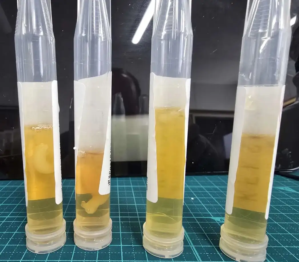
According to the Twitter user Janiesaysyay, the photo was proof that living people are growing calamari clots: [https://

Peter Daszak posted this tweet: [https://

When I explained to Daszak how McCairn's immunoadsorption circuit is unable to even remove the microclots, Daszak retweeted my tweet and told me "I'm glad you're exposing this horrible grift":
Then Daszak blocked me for some unknown reason. Black Tom said that I was in cahoots with Daszak, but when I showed that I had already been blocked by my new friend Pete, Black Tom said we were covering up our tracks: [https://



Jikky said that me and Daszak are affiliated with the same agency: [https://

These were the types of replies Daszak received from McCairn's fans: [https://



One user on Twitter said that McCairn was threatening Peter Daszak's "evil harvest": [https://

Another user suggested that Daszak had organized the attacks against McCairn: [https://

Janiesaysyay said that Daszak had inflicted an extinction-level event on humanity: [https://

In August 2026 McCairn's patient Robert Benson posted a video where he said it had now been about 4 months since he returned from Japan, and he said: "So coming out of Japan, I was 80, 90 percent. I felt my usual self, I was doing real - doing really well, and that carried through, right, you know, into July. I'd say mid-to-late July, all of a sudden I could kind of feel things coming back. It felt like I was had a - you know, wet blanket thrown over my brain again, so I thought things might be coming back. I did go get my spike proteins tested - and my spike protein antibodies tested - I was over 25,000 again. So those numbers come back high." [https://
So mid-July would've been about 3 months after the treatment. Based on how the symptoms of autoimmune conditions rebound within weeks after immunoadsorption treatment, I would've expected the effect of McCairn's treatment to last an even shorter time. The Sinovac victims guy also said that his symptoms had rebounded by four days after the DFPP treatment.
I don't know if McCairn's other patients have experienced a faster or slower rebound than Robert Benson, or no rebound at all.
A few days later Robert Benson posted another update, where he now said that he had tried therapeutic plasma exchange in the United States. He said most of his issues were cognitive and mental, and while the gains from TPE did not last from a mental standpoint, the TPE fixed his gut issues, and it fixed a rash in his foot within days from the treatment. But he said that the catheter was hooked up to his arm in the TPE, and he speculated that perhaps the treatment in Japan had a bigger cognitive benefit because he said that "I think that actually what happens in Japan when they go through the jugular, and use the catherer, that I think it's better allowed to pull the clots out of my brain, that cause me the - just my, numbness in my daily life, every day. So I think that's a big factor." [https://
McCairn should stop falsely advertising his protocol by claiming that the jugular catheter somehow helps to suck out toxins from the brain, or to suck out clots from the brain. He should also inform his patients that his protocol likely does not remove microclots in the first place, but that TPE might remove microclots when a centrifuge is used to separate the plasma.
McCairn's fan Janiesaysyay is now going around Twitter promoting the Edogawa protocol as a treatment for removing calamari clots: [https://
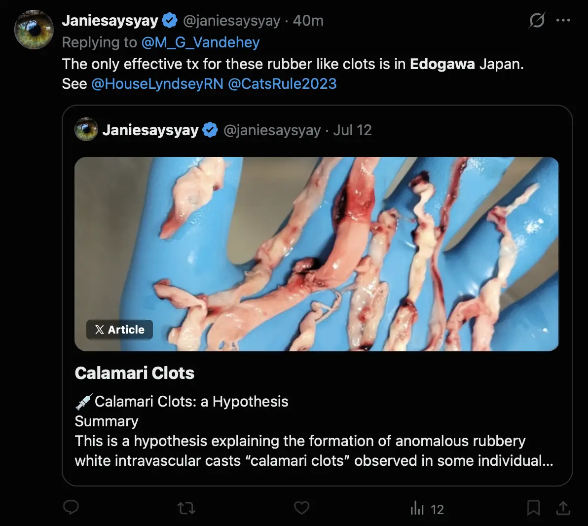
Earlier one of McCairn's patients showed a photo of a clot in her plasma, and she said the photo was undeniable proof that calamari clots are occurring in living people: [https://

Janiesaysyay also claimed that the patient had calamari clots coming out of her blood: [https://

McCairn has said that he thinks the calamari clots are a post-mortem phenomenon, because he said that if the clots would occur in living people, the clots would be reported by cath labs, and he said he had heard from the staff of a Japanese hospital that they were not seeing calamari clots in living people. [https://
In August 2026, Doug Kell and Resia Pretorius posted a preprint titled "Atherosclerosis as a Localised Amyloid and Proteostasis Disorder". [https://
I had already informed Doug Kell earlier that AmyloGram is meant to be used with peptides and not proteins, and proteins virtually always get an AmyloGram score above 0.5. I had also showed him how the AmyloGram score is simply the maximum score of 6 aa segments within the input, so the score is not a meaningful measure of the overall amyloidogenicity of a whole protein. But in his preprint he didn't mention how the AmyloGram score is calculated, or why full proteins are almost always classified as amyloidogenic by AmyloGram.
The AmyloGram score centers around 0.5 for random 20 aa peptides, because the score is calibrated to be meaningful for peptides but not for proteins. But the average score is about 0.8 for random 500 aa proteins, because random 500 aa proteins virtually always contain nonzero 6 aa segments that are classified as amyloidogenic:
library(AmyloGram); library( biogram); data( AmyloGram_ model) chars=strsplit( " A C D E F G H I K L M N P Q R S T V W Y ", " ")[[ 1]] # average score of 100 random 20 aa sequences l=replicate(100, paste( sample( chars, 20, T)), F) l=setNames( l, 1: length( l)) mean( predict( AmyloGram_ model, l)$ Prob) # 0. #4709998 average score of 100 random 500 aa sequences l=replicate(100, paste( sample( chars, 500, T)), F) l=setNames( l, 1: length( l)) mean( predict( AmyloGram_ model, l)$ Prob) # 0. 8364628
Here when I ran AmyloGram for random amino acid sequences of each length between 6 and 1,000 aa, all sequences longer than 150 aa got a score greater than 0.5, which is the default cutoff AmyloGram employs to determine if an input sequence is likely to be amyloidogenic or not: [clot4.
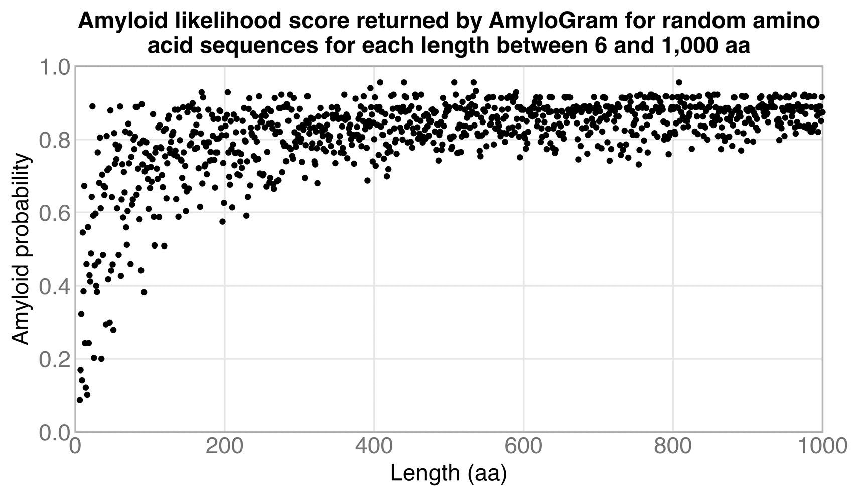
The paper about AmyloGram said that AmyloGram was trained on a set of peptides with a length between 6 and 25 aa, and a benchmark of AmyloGram relied on input sequences with a length between 6 and 14 aa. [https://
The same problem is much worse for a protein with a length of hundreds of amino acids, so if even one of the hundreds of 6 aa segments gets a score above 0.5, then the entire protein gets classified as amyloidogenic.
In the new preprint, Kell and Pretorius wrote: "Langley and colleagues (Langley et al. 2017) sought biomarkers of high-risk atherosclerotic plaques, and identified a 4-biomarker signature (matrix metalloproteinase 9, S100A8/S100A9 (calprotectin), cathepsin D, and galectin-3-binding protein). Their AmyloGram scores are respectively 0.9148, 0.7768 (for S100A8), 0.7941, and 0.8661. Clearly, each is highly amyloidogenic ...". But scores of around 0.8 or 0.9 are typical for whole proteins, so the scores are not evidence that the proteins are highly amyloidogenic.
Kell and Pretorius also wrote: "a computational analysis based on AmyloGram showed the strongly amyloidogenic potential of proteins found in various atherosclerotic plaques". But that's wrong, because a high AmyloGram score is not evidence of a protein having a "strongly amyloidogenic potential".
In the new preprint, Kell and Pretorius wrote: "Thus, while 79% of human proteins have an AmyloGram score exceeding 0.7, with a median value of 0.81, typically more than 90% of proteins in plaques have AmyloGram scores exceeding 0.7, with a median score around 0.86. The probability-of-superiority values are around 0.615-0.627 against the human proteome. In addition, a Mann-Whitney test bootstrapped 20,000 times gave a 95% CI 0.0411-0.0497 for the difference in median AmyloGram score in Lewy bodies vs the human proteome."
So the difference in the average AmyloGram score was only about 0.05 between the two sets of proteins. I thought the difference might have been explained by a different length distribution between the sets.
Therefore I downloaded both Kell's set of 1,459 proteins associated with plaques, and I downloaded a reference set of human proteins from UniProt similar to Kell's reference set. [https://
Then when I divided the proteins to 10 length strata and I calculated the average AmyloGram score within each stratum, the set of proteins associated with plaques got a lower score within the 4 shortest strata, and the set of reviewed human proteins got a higher score within the 6 longest strata:
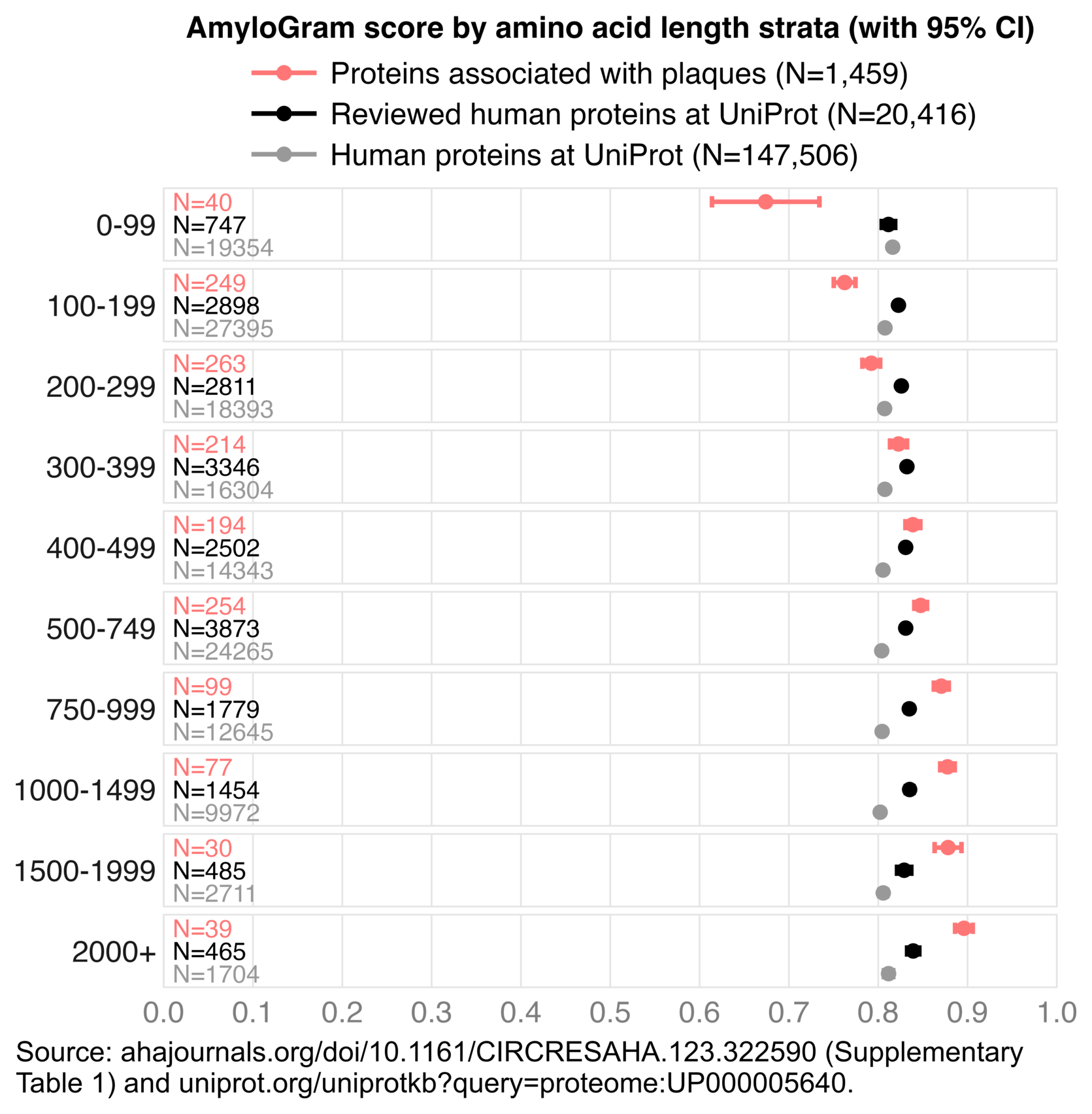
I don't know what explains the discrepancy between the short and long proteins. But regardless, the results still show that proteins associated with plaques do not consistently get higher AmyloGram scores than the reference proteins.
I showed the length-stratified results to Doug Kell, and I informed him of how the AmyloGram scores are not meaningful for proteins but only peptides. [https://
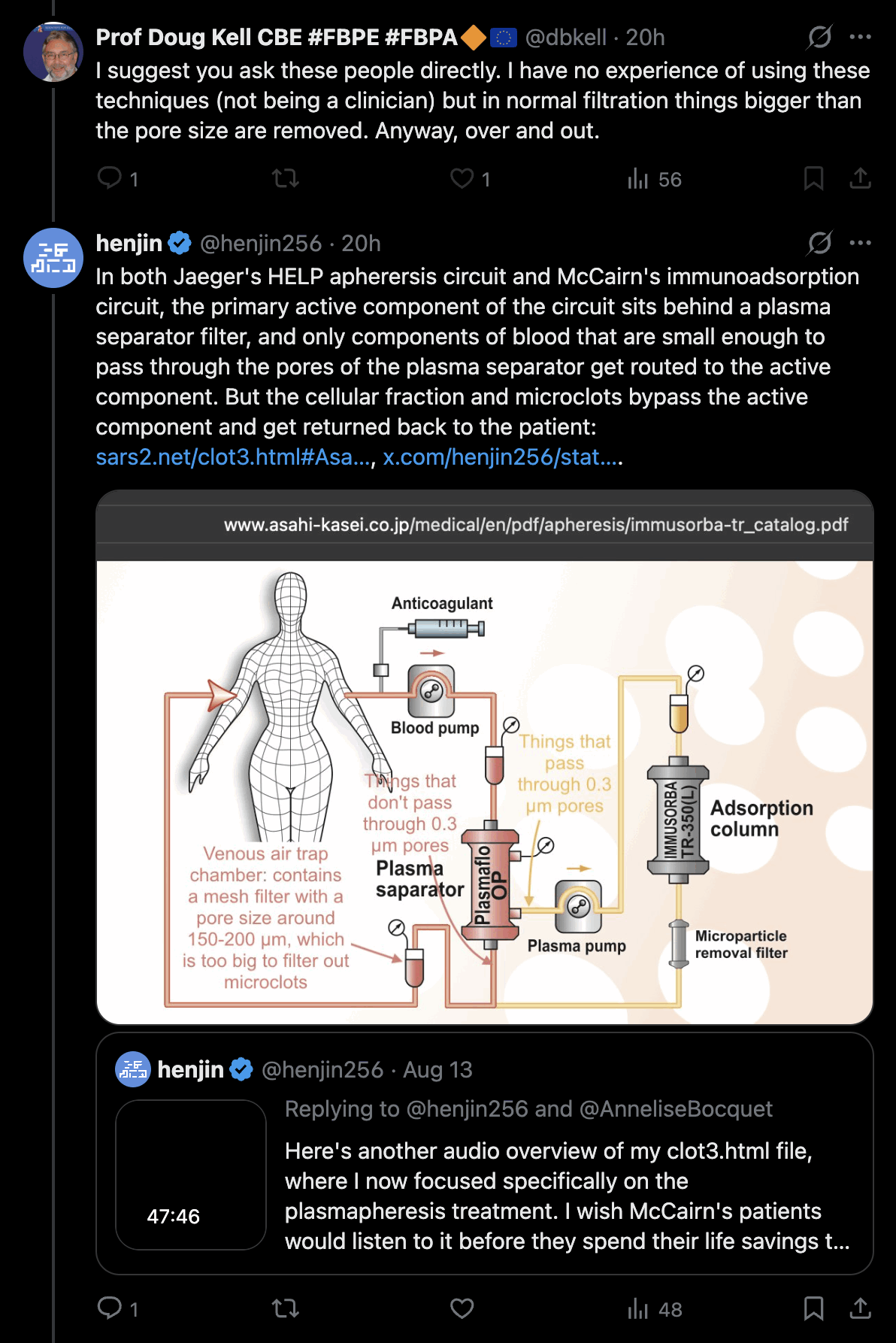


I had just helped Kell identify a fundamental problem in his recent preprint, so he should've at least thanked me for helping him, so he'll know to correct the preprint before it gets published in a journal. Or he should probably withdraw the preprint entirely, because the primary finding of his preprint was not valid. But instead he just said that I was a "disaffected troublemaker" who was "off on an ego trip".
I understand if Kell and Pretorius don't want to get involved in the debate about McCairn's protocol, because it's such a radioactive topic. But on the other hand their papers serve as a key device McCairn employs to make his patients afraid of the microclots, and to trick his patients into thinking they have to give up their life savings to get rid of the clots. So once McCairn's grift comes crashing down, it might also have an adverse effect on the reputation of Kell and Pretorius (especially since Pretorius has earlier supported Beate Jaeger's HELP apheresis as a potential method to remove the clots, even though Jaeger's HELP protocol seems to have the same pore size problem as McCairn's immunoadsorption protocol).
The website of the apheresis center at Cyprus says: "Please note that we currently no longer utilize traditional Immunoadsorption as part of our routine clinical protocols. Over time, the Apheresis Center has transitioned toward using Inuspheresis® as our preferred and selected form of Immunoapheresis within our broader Combination Therapy Approach." [https://
The website also says:
Despite initial studies showing no effect for Immunoadsorption in Long Covid, clinical experience tells a different story. While not as effective as H.E.L.P. Apheresis, Immunoadsorption has shown a 10-30% success rate in clinical settings, compared to the 80% success rate of H.E.L.P. Apheresis. The results of Immunoadsorption are not always permanent, potentially due to its single mode of action and the high burden of fast application.
Further research, such as this study:(https://
www. ), is needed to improve success rates. New adsorbers, combination treatments, and different application schemes may enhance effectiveness.medrxiv. org/ content/ 10. 1101/ 2024. 06. 18. 24309100v1 All three methods - Immunoadsorption, H.E.L.P. Apheresis, and Inuspheresis® - belong to the wider family of Immunoapheresis and Therapeutic Apheresis when used to target pathogenic autoantibodies and inflammatory mediators.
At our center, we continue to evolve our approach based on patient outcomes, scientific developments, and the complexity of chronic inflammatory conditions. While Immunoadsorption remains a recognized therapy, we increasingly utilize broader therapeutic approaches such as Inuspheresis® and combination therapy protocols to address multiple overlapping disease mechanisms in a more comprehensive way.
The apheresis center at Cyprus might be the only provider of apheresis treatment that has comprehensive experience providing multiple types of apheresis treatment to long COVID patients. For example Edogawa seems to only provide IA treatment, Jaeger provides only HELP apheresis, and some American centers only provide TPE, but the center at Cyprus might be uniquely positioned to compare the efficacy of different forms of apheresis for long COVID (even though I don't know if the center has collected any rigorous numeric measurements to compare the efficacy, and the center didn't explain how they determined that immunoadsorption had a "10-30% success rate" and HELP apheresis had an "80% success rate").
A blog post by the Cyprus center said that HELP apheresis was "by far the most successful type of apheresis we use", and the post said: [https://
Immunoadsorption does one thing only, but at a very high efficiency: Removing autoantibodies. From our clinical experience it is only indicated if the patient has certain very high autoantibody levels. If applied too fast (the often standard 6 sessions in 2 weeks is what we believe (from anecdotes of patients who did immunoadsorption at higher speed as we apply) is too fast for many patients) there is a high risk of 'crashing' or getting worse, sometimes even permanently. Another issue is that most results are only permanent for approximately a third of the patients that undergo the treatment.
In general we have had better results with IVIG treatment so far than with Immunoadsorption - but both are a good combination with H.E.L.P. if applied correctly. More research is needed in this area. H.E.L.P. removes autoantibodies too (at a lower pace without creating issues) and outperforms Immunoadsorption in all other areas, another reason why it is the preferred first line treatment of our medical team.
In August 2026, McCairn was interviewed by Michael Gray Griffith and Paul Oosterhuis on a channel called Cafe Locked Out. [https://
Two months earlier Cafe Locked Out interviewed an Arcturian guy called IRa, who says he is an emissary of the Galactic High Council of Planets, and he says his mission is to assist humanity to ascend from the third dimension to the fifth dimension: [https://

IRa the Arcturian also says that vaccines contain graphene and nanobots: [https://


Next Cafe Locked Out interviewed Greg Harrison, who is possibly an even more ridiculous person than IRa the Arcturian. So I thought there was no person more ridiculous who the channel could've interviewed next, but then they interviewed Kevin McCairn.
During the two months before the interview, me and other people on Twitter had figured out that McCairn's immunoadsorption treatment is unlikely to remove microclots from the blood, because the clots are too big to pass through the 0.3 µm pores of the plasma separator. But McCairn had avoided addressing the pore size problem on Twitter, and he had tried to sweep the problem under the rug. He also hadn't done new livestreams or interviews in a long time, so I thought that now during the interview with Cafe Locked Out, he would finally address the pore size problem, but he never did.
Me, Allen Green, and Markus have also said that it was misleading how the treatment at Edogawa was being marketed as "DFPA", because if treatment labeled DFPA is not available outside of Japan, then McCairn's patients will think that the treatment relies on some kind of unique technology that is not available outside of Japan. But in reality DFPA appears to be a term invented by Edogawa, and what Edogawa calls DFPA is just regular immunoadsorption, and similar immunoadsorption treatment is available at a lower cost in other countries.
I don't remember McCairn having earlier referred to the treatment as immunoadsorption, but now at time 10:07, he said: "So we do a procedure called - it's immunoadsorption apheresis - but we're sort of going by dual filtration plasma apheresis, because it's kind of a dual filtration setup, um, but with the immune adsorption."
I used to think that the acronym DFPA stood for "dual filtration plasma adsorption", because the acronym "PA" stands for plasma adsorption in Japan. And also the term "plasma apheresis" as two words is less common than "plasmapheresis" as a single word, and "plasmapheresis" is abbreviated as PP and not PA in the acronym DFPP.
But generally in contexts related to the Edogawa protocol, the term DFPA seems to be more commonly defined as "dual filtration plasma apheresis" than "dual filtration plasma adsorption". The only users on Twitter who I found using the latter definition were me and McCairn's patient Burning_mama47. [https://
So apparently the letter A in DFPA is supposed to stand for apheresis and not adsorption. Which is really confusing, because then the term DFPA ("dual filtration plasma apheresis") is nearly the same as DFPP ("dual filtration plasmapheresis"), but with plasmapheresis written in a different way, even though DFPP is not the same as the IA treatment that is confusingly called DFPA by Edogawa.
The edogawadfpa.com website says that Edogawa's "clinical program is centered on Dual Filtration Plasmapheresis, also described as DFPP or DFPA in patient materials". [https://
At time 23:44, Griffith said: "Kevin's - and he's in Japan - and he is working on treatments to remove the spike protein and the amyloid - we call them calamari clots - but the amyloid clots from people's blood." At that point McCairn should've said that the Edogawa protocol shouldn't be advertised as a method to remove calamari clots from the blood, because he doesn't even know if the so-called amyloid microclots are related to calamari clots, or if calamari clots occur in the blood of living people, or if calamari clots are even real. And McCairn should've said that his immunoadsorption treatment likely does not even remove microclots from the blood in the first place, because the clots are too big to pass through the pores of the plasma separator. But McCairn didn't say any of those things, and he didn't seem to have a problem with Griffith conflating microclots with calamari clots.
I think it's highly deceptive for McCairn to advertise his protocol as a method to remove microclots, and even more deceptive to advertise his protocol as a method to remove calamari clots, because calamari clots are not even real.
At time 26:15, McCairn said: "When I receive slides from people, I have to check the glass, right, just to see how much it's sort of picked up contaminant. Because it - loads of things glow in UV, right. If you just sort of think about, you know, you try and look sharp at the rave, and the UV lights come on, and basically all of your dandruff lights up like, uh, like a, you know, disco ball, right? And so yeah, it's very important to be able to - well, everything I say, just like, if - if it's come through the post, and I have to say, 'Look, I have to caveat your results with the quality of the slide that you've made.' I don't expect people to have a clean room and lab-grade slides coming through. And just to tell you how sort of tricky the methods are, right - which is, uh, I don't have a box here, but, um, I would tell people to get these slides. Uh, hang on."
Then he went to pick a box of lab-grade slides, which he said were pre-cleaned slides that cost 400 bucks a box, and he said there's a contamination risk if his followers buy cheaper slides that are not pre-cleaned.
But I don't think the main contamination risk comes from the slides being contaminated right out of the box, but rather from dust falling on the slide while the blood is drying, or when McCairn looks at the slide in Japan, or at some point in between.
So his followers not having pre-cleaned slides was a weird explanation for why the slides would be contaminated.
Then at time 29:37, McCairn said: "There's always people's - just environment, et cetera - and, um, it's not the cleanest of tests. But I would add that actually, I think it's one of the better tests even compared to pulling IV blood. And the reason for that is that what we see presenting is basically small vessel, small fiber disease. And it's a spike-driven angiopathy. And the lancet test on the finger - where you're basically going into, um, highly vascularized, highly innervated tissue, and squeezing out a drop of blood from there - is actually, it's actually - I see more hits of that than pulling from an IV line or the central line. And that's because I think - what I think is happening is the amyloid is just sort of coating those small vessels, a bit like sort of paint in a way. And they sort of build up, they come a bit rigid."
So apparently McCairn has tried analyzing samples of blood taken directly from an IV line or central line, but he found that the samples didn't contain as many of the so-called microclots as the samples of blood he received in the mail. I think it's because he misidentified dust as microclots in the samples he received in the mail, and the blood taken straight from an IV line didn't contain dust.
At time 1:00:31, Griffith said about the leaders of Australia: "We became the Jews overnight, and you guys became Nazis overnight. And you fucked up a whole country." But McCairn didn't push back against what Griffith said, even though earlier McCairn has pretended to be opposed to the Jews. There was even a moderator on his Discord who was a member of the neo-Nazi group Atomwaffen. But I guess now McCairn has realized that there's more money be extracted from anti-vaxxers than from antisemites, so he won't stand up against someone who presents the Jews in a favorable light and Nazis in an unfavorable light (even though I think it's fairly likely that McCairn was always a fake antisemite like Stew Peters, who was never sincerely opposed to the Jews, but who covertly worked for the Jews in order to make antisemites look ridiculous).
At time 1:18:55, McCairn spoke of a "rag bag of indigents who were just buying into graphene oxide and nanotech in the vials", and he said "a lot of that was sort of controlled psychological operations". But Griffith failed to mention how he had recently interviewed an Arcturian emissary from the High Council of Planets, who says that vaccines contain graphene and nanobots. And Griffith didn't say that when he did a video about nanobots with David Nixon and Mateo Taylor, Griffith presented a video of Alex Jones being scanned with a nanobot scanner as evidence that people are now filled with nanobots. [https://
At time 2:23:34, Griffith said: "If you want blood analysis done, it's there, over here now. We often work with a guy called David Nixon as well. He does blood analysis. He can't tell vaccinated blood from unvaccinated blood anymore. He claims it looks the same." Then to McCairn's credit, he said that David Nixon is "somewhat nanobot-tarded". But I guess if McCairn saw there was more money to be extracted from the nanobot guys than from the anti-vaxxers, he would say that people are now infested with nanobots, and the only way people can get rid of the nanobots is to give him their life savings.
At time 2:30:18, McCairn said: "So this is the system that we're using. It's made by a company called Asahi Kasei. It's Immusorba TR-350. It's specific for neuropathy. And it was the one thing that I, um, heed in on, when we were sort of trying to pick out the therapy. And, you know, I was just was like passing me this list as we're thinking about patient N2. And I've got the symptom profile - so I'm looking at him, it's coming through - and I see this one. And it's for, uh, neuropathy, um, what do they call it, CIDP, right, uh, chronic inflammatory demyelinating polyneuropathy, and Bell's palsy they use it for, and myasthenia gravis. And I was like, 'Choose that one. Grab the autoantibodies for that.' And that's what this one is doing."
So was McCairn personally involved in choosing immunoadsorption as the preferred modality of plasmapheresis? He made it seem like he was browsing through a list of different plasmapheresis modalities, and he arbitrarily decided to pick immunoadsorption, because he guessed it might have treated the symptoms of one of his first patients.
But was McCairn even aware of how the center in Cyprus says that they found immunoadsorption to be less effective in treating long COVID than DFPP or HELP? And since McCairn primarily advertises his plasmapheresis treatment as a method to remove microclots, did he somehow evaluate if immunoadsorption was more likely to remove microclots than other forms of plasmapheresis?
McCairn makes it seem like the form of plasmapheresis provided at Edogawa is somehow superior to other forms of plasmapheresis, even though he seems to have selected immunoadsorption as the preferred mode of plasmapherersis more or less arbitrarily, and he probably doesn't even know if immunoadsorption is more effective at treating long COVID than DFPP or HELP or TPE.
But in any case, it seems highly irresponsible for Edogawa Hospital to allow someone like McCairn to have this much involvement in the treatment. McCairn is a research scientist and not a clinician, so I think according to Japanese law, McCairn shouldn't even be allowed to perform diagnostics as part of the treatment protocol, or to direct patients to get the treatment.
About two weeks after McCairn's patient Kelsey Shields had returned home from Japan, she said she felt a reduction in brain fog and an increase in energy, but she said that "I still have a very limited energy envelope, significant POTS/orthostatic intolerance and ME/CFS, must be extremely careful about overdoing it, and still rely heavily on caregivers." [https://
She also said: "There have also been scary new medical issues since returning home: intermittent numbness and tingling in my hands and feet and, most recently, acute urinary retention - my bladder stopped emptying properly. I currently have an indwelling catheter and am undergoing further neurological/urological evaluation."
And she said: "Some earlier patients have reportedly experienced decline around the 4-6 month mark, while others have maintained improvements longer."
In another tweet she wrote she had jugular venous stenosis, and she posted a response by an AI that said: "MRV imaging demonstrates significant bilateral jugular venous stenosis, particularly severe on the right side, with compensatory vertebral venous outflow development. This finding correlates with patient's symptoms of headaches, pressure behind eyes, neck stiffness, and pain at the base of skull. The stenosis appears to worsen as the jugular veins descend from the head." [https://
I don't know if the jugular stenosis was caused by the plasmapheresis treatment, because Gemini said:
Jugular venous stenosis can occur as a delayed complication of plasmapheresis when large-bore central venous catheters are inserted into the internal jugular vein. Although plasmapheresis itself - the mechanical removal and exchange of blood plasma - does not alter the physical structure of the vessel, the invasive placement and presence of the jugular catheter can directly cause vascular trauma and subsequent narrowing.
The primary mechanism behind catheter-induced stenosis involves mechanical injury to the delicate endothelial lining of the vein. Inserting the catheter, movement of the stiff tubing against the vessel wall, or turbulence created by high-flow apheresis can trigger localized inflammation, intimal hyperplasia, and smooth muscle proliferation. Additionally, catheters can promote localized thrombus formation, which may organize over time into fibrotic tissue that permanently restricts the vein's inner diameter.
Risk factors that elevate the likelihood of developing internal jugular stenosis include prolonged catheter dwell time, larger catheter lumen sizes, multiple insertion attempts, underlying catheter-related infections, and a prior history of central venous line placements. While many cases of jugular stenosis remain asymptomatic due to collateral venous drainage, significant narrowing can lead to symptoms such as facial or neck swelling, localized discomfort, or persistent difficulty obtaining adequate blood flow during subsequent apheresis or intravenous procedures.
In September 2026 when someone said that McCairn needed to do a sham-controlled study for his treatment, McCairn replied "Hard to sham control central line access", even though he knew that the IAMPOCO trial employed a jugular central line, but the trial was still sham-controlled: [https://
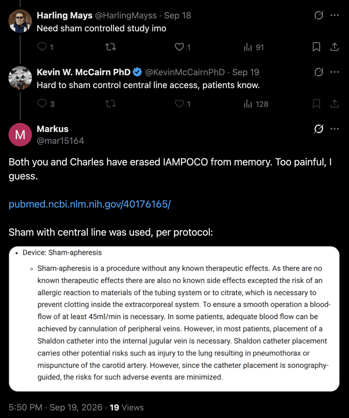
Rixey didn't say anything about the IAMPOCO trial either: [https://

A couple of days later McCairn again said "Hard to sham a central line going into the Jugular", even though Markus had just reminded McCairn about how the IAMPOCO trial was sham-controlled: [https://

In October 2026, McCairn did a Twitter space with Nurse Michele, who has a podcast on America Out Loud Network. [https://
At time 1:28:41, McCairn said: "There was this 19 nucleotide insert, which basically, um, imbued the spike protein with the ability to be cut by an enzyme in the body called furin." But of course the FCS insert is only 12 bases long, but 19 bases is the length of the BLAST match to a Moderna patent that contained the 12-base insert and 7 bases before the insert. And the match to the Moderna patent was by chance, and if you do a BLAST search of the whole genome of SARS-CoV-2 against patent sequences published before 2020 that match the keyword "Moderna", the 19-base match gets an E-value of 381, and there are hundreds of other similarly close or closer matches. [fleming.
At time 3:05:54, McCairn said: "The only new thing about this therapy is the combination of therapies. So this approach, and using dual filtration plasma apheresis and immune adsorption, has been in place in Japan for thirty - more than thirty years." I don't know if by "dual filtration plasma apheresis" he meant DFPP (which stands for "dual filtration plasmapheresis", where "plasmapheresis" is usually written as a single word) or DFPA (which is usually defined as "dual filtration plasma apheresis" but sometimes "dual filtration plasma adsorption"). But in either case, it's confusing how people keep referring to the treatment at Edogawa as DFPA or DFPP, even though in reality the treatment seems to just be immunoadsorption.
At time 3:14:50, McCairn said: "That thing going in your neck, right, that's a central line going in. It's not - this isn't a health spa EBOO treatment, right. And so, unfortunately we've had two adverse events from that. Which is basically just a bleed into the thoracic space." So he seems to have now had a second patient with hemothorax, apart from AprilWW911LC who had a hemothorax in July 2026. [mccairn.
At time 3:20:04, McCairn said: "The therapy is available in the US, if you're in the US military and you get sepsis on the battlefield, you can get dual filtration plasma apheresis. And there are numerous - how would you say - people who are putting forward - how should we say - technology around filtration that isn't being - allowed to be released to the American public. Now the thing is - I was thinking about this - this might even be due to the fact that there are - the Japanese have maybe tied up all this tech in patents, as well as all their immunoabsorption technology, right. So it sort of occurred to me the other day that they can't do it. So they can use it - that they can get away with it. So DARPA spent billions on, one, trying to develop artificial blood, right, that's room temperature stable and can be kept for a while, a long time. And the other is how to treat sepsis on the battlefield."
He made it seem like the TR-350 immunoadsorption column used at Edogawa was some kind of secret Japanese technology, because the column is not licensed in the United States, even though the column is licensed in many European countries, and it has even been used to treat long COVID in Germany.
I think what McCairn meant by the US military technology is the CytoSorb hemoadsorption device, which is developed by a company called CytoSorbents. But CytoSorb is neither DFPP or immunoadsorption. CytoSorb doesn't even include a step of separating plasma from the blood cells, which is the function of the first filter in DFPP, so it's highly misleading for McCairn to refer to CytoSorb as "dual filtration plasma apheresis". I believe CytoSorb is not even used by the US military on the battlefield, but CytoSorbents has simply received funding from the US military and DARPA, so McCairn portrays CytoSorb as some kind of secret military technology, even though in reality it's primarily used in the civilian field. [https://
Gemini said:
The U.S. military does not use the standard CytoSorb column as part of its routine field medical care or active-duty clinical inventory, primarily because the device lacks full standard approval from the U.S. Food and Drug Administration (FDA). Standard deployment of medical technologies within U.S. military operations generally requires broad FDA clearance or approval.
However, the U.S. Department of Defense (DoD), the U.S. Army, and the Defense Advanced Research Projects Agency (DARPA) have maintained significant research and development partnerships with CytoSorbents Corporation for many years. Millions of dollars in military research contracts have been awarded to evaluate and adapt the company's core bead-polymer adsorption technology for combat casualty care, trauma management, severe burn treatment, and organ preservation.
At time 3:37:28, McCairn said: "I was like looking through, and there's like lists of all these different treatments that are available, with respect to the canister you can get. And then I go through, and then there's the neurological section, and then I - there's the canister Immusorba, for chronic demyelinating - CIDP it's called - chronic inflammatory demyelinating pathology or syndrome, right - and that's one of the predominant symptoms that we see. And it responds very very well to this treatment." Actually the letter P in CIDP stands for polyradiculoneuropathy. But anyway, McCairn continued: "And why is that? Well, two. For one is it removes the aggregates - the immune aggregates - that come from the disseminated intravascular coagulopathy, which is essentially the microclotting. The immunoadsorption, which is the - it's tuned to take, or absorb out - autoantibodies that are teeing up and attacking the peripheral and central nervous system."
McCairn has earlier used the term "aggregate" as a synonym for microclots, so what does he now mean by immune aggregates? He now seems to have said the "immune aggregates" are essentially the same as "microclotting". But he already knows that the microclots are too big to pass through the 0.3 µm pores of the plasma separator, so the microclots never even pass through the immunoadsorption column.
Both during this interview and the earlier interview with Michael Gray Griffith, McCairn seems to have now started referring to his treatment as immunoadsorption (even though he still sometimes confusingly refers to the treatment as "dual filtration plasma apheresis"). But now he seems to have also invented a new term for the microclots, which he now calls "immune aggregates", as if adding the word "immune" to the name of the microclots would somehow mean that the clots would be removed by immunoadsorption.
At time 3:45:25, McCairn now said he had started collecting numeric measurements of the efficacy of his treatment, so that he's now doing a questionnaire where he asks his patients each week how they were feeling. He said: "The questionnaire is sort of like, 30-odd questions covering these three areas. And, so, basically this has just been normalized to zero. And the patient just rates 1 to 10, right, how severe they are. And so, the first marker on this is them at baseline before treatment, and then you can see the following four treatments. And then the last one here is a few that have gone out extended, and it's mixed over different distances. But, um, the p - so the probability of it being to chance that this therapeutic level is so enormously, enormously successful, and the p-score is so low that it's - you're getting into odds where, you know, they start talking about, I don't know, you know, like quantum field type, you know - that sort of level of statistical strength." But his questionnaire was not controlled for a placebo effect, and McCairn's patients have been led to believe that his treatment is some kind of a miracle cure that is supposed to make them feel amazing. In the German IAMPOCO study that compared TR-350 against a sham treatment, there was no significant difference in subjective wellbeing between the treatment group and sham group. [https://
At time 4:00:20, McCairn said: "Everything's being pushed through a filter. Everything is reduced, right. Cytokines are reduced, the aggregates are reduced." By aggregates he probably meant microclots, even though he already knows that the microclots are too big to pass through the pores of the plasma separator, so the microclots don't get pushed through the immunoadsorption column.
At time 4:14:12, McCairn said: "Because of the, uh, predilection of these autoantibodies attacking central nervous system, by going into the jugular vein, basically you're targeting that outflow of the brain, right? Because it's all - it all has to go - cranial circulation basically goes down via the jugular vein into the heart, okay. And so the concentration of autoantibodies - so if that's your cylinder, right, if you're trying to pull against the whole of the body through a small peripheral vein, and trying to - and you've got a fixed volume, through which to expose through autoantibodies right? Which is going to work better: this one is pulling up at the arm, or the one where it's strapped, right, or pulling right from the neck, and the cranial circulation, right? So it's literally taking the most concentrated form of brain waste, and cleaning it, rather than trying to clean the diluted form that keeps getting moved around in circulation."
But I don't think he has shown that the blood vessels of the brain would somehow have an elevated concentration of autoantibodies relative to other parts of the circulatory system. And even if the blood vessels of the brain temporarily had an elevated concentration of autoantibodies, the autoantibodies would soon diffuse out to other parts of the circulatory system.
Grok said:
McCairn has not demonstrated, and standard physiology does not support, a meaningfully elevated concentration of free autoantibodies in the venous outflow of the brain relative to the rest of the circulatory system. Even a temporary local difference would be irrelevant because of rapid redistribution.
In diseases involving autoantibodies against CNS targets, such as certain autoimmune encephalitides or NMOSD involving AQP4, the bulk of pathogenic antibody production is peripheral. Autoreactive B cells and plasma cells reside mainly in secondary lymphoid tissues including lymph nodes, spleen, and bone marrow, and they release antibodies into the systemic circulation. These circulating antibodies can enter the CNS if the blood-brain barrier is compromised. In some conditions there is also intrathecal synthesis, in which B cells that have migrated into the CNS produce antibodies locally in the CSF or parenchyma. This is why CSF can sometimes be more sensitive than serum for certain antibodies, notably NMDAR, while for others, notably AQP4, serum is more sensitive and CSF positivity largely tracks high serum titers plus barrier leakage. Absolute antibody levels are still typically far higher in serum than in CSF because the total immunoglobulin pool is vastly larger in blood.
None of this creates a sustained higher concentration of free circulating autoantibodies in the internal jugular vein compared with, say, an arm vein. Jugular blood is simply venous drainage from the head and brain mixed with blood from other cranial structures. Any free antibody leaving the CNS compartment into that venous blood is a tiny fraction of the total systemic pool and is immediately diluted and mixed.
The circulatory system mixes thoroughly and quickly. Blood from the jugular veins returns to the right heart, is pumped through the lungs, and is distributed systemically. Any transient local elevation in one venous territory is short-lived and does not persist as a therapeutically useful concentration gradient. Once antibodies are in the plasma compartment, their concentration equalizes across the circulating blood volume on a timescale much faster than an apheresis session.
Plasmapheresis or immunoadsorption removes a portion of the systemic circulating plasma antibodies, typically aiming to process roughly one to one-and-a-half plasma volumes per session, repeated over days. Efficacy depends on how much of the circulating pool you process and how much redistributes from extravascular spaces afterward, not on whether you withdraw the blood from a neck vein versus an arm vein. The access site is chosen for flow rate, stability, and practicality.
There is research showing modestly higher levels of certain brain-derived proteins, such as some Alzheimer's biomarkers including Aβ peptides or p-tau, in internal jugular vein blood versus peripheral venous blood. That is a different phenomenon involving proteins produced in the brain and draining outward, and the differences are relatively small. It does not extend to free autoantibodies in a way that would make jugular access superior for antibody removal.
In short, the premise that the jugular catheter is selectively pulling the most concentrated form of brain waste lacks supporting evidence and conflicts with how antibodies are produced, distributed, and mixed in the body.
Grok also said:
Brain toxins and waste are cleared mainly through the glymphatic system and meningeal lymphatic vessels, not primarily through the jugular veins.
CSF enters the brain along spaces around arteries, mixes with interstitial fluid (aided by aquaporin-4 channels on astrocytes), and carries dissolved waste out along spaces around veins. From the surface of the brain, meningeal lymphatic vessels in the dura then drain this fluid, along with solutes and waste products, to the deep cervical lymph nodes. The lymph eventually returns to the bloodstream.
The internal jugular veins mainly carry deoxygenated blood from the brain's venous sinuses back to the heart. While some soluble waste can enter the blood, the glymphatic-meningeal lymphatic route is the main dedicated pathway for clearing larger molecules and metabolic byproducts from the brain.
At time 3:27:17, McCairn said: "The sneaky thing is - what they do is they use replacement population to hide the all-cause mortality rates, right. So all the time you're bringing in, like foreign workers, which is happening across Europe and across the US, right, your sort of, baseline population stays the same or is increasing, right. And so in a sense it camouflages the increased mortality that you would see, were there - were the population remaining the same prior to it being COVID." I guess he means that if excess mortality was calculated based on a crude mortality rate that was adjusted for population size but not for age, then the migrants would be mostly working age people who would have few deaths, so the migrants would inflate the denominator for population size but not the numerator for deaths. But reasonable methods of calculating excess mortality are adjusted for age, and not only population size. And in the United States and European countries, there is now low excess mortality even if you adjust the calculation both age and population size, and not only population size. So McCairn's explanation doesn't make sense.
Then next Nurse Michele said: "Well honestly, besides yourself, John Beaudoin, maybe the Ethical Skeptic, and a few others that are out there, who's even saying that?" Then McCairn said: "Not many. And shout out to both those gentlemen. Um, I think they - I think they've both done amazing work." I have shown McCairn many times how Ethical Skeptic has falsified the data in his plots, so why would McCairn continue to say that ES does amazing work? Last year McCairn promoted a plot where Ethical Skeptic claimed that ages 0-4 had 77% excess deaths in early 2025, so I had a long exchange with McCairn where I clearly showed him how Ethical Skeptic fabricated the data in his plot, and McCairn even did a series of three streams about the exchange. clot3.
At time 4:09:14, McCairn said: "If we know spike protein causes abnormal clotting, why are we surprised that we see in patients, one, a propensity for when - because it's not coming out like that, right? Whilst it's warm and in the patient, it's not physically [...] the body is keeping it stabilized. But like any fluid, right, if you pull it out, and you don't, um, actively pull, uh, so you use, um, citrate and EDTA to sort of pull, uh, the factors out for initiating the clotting cascade, and so, um, the - of course, in that situation, it will clot. It will - doesn't take much temperature change, bang it against the side of the freezer, et cetera. And nearly every patient's first pre-filtration, they seem hyper-coagulable. Whereas by the end of the treatment, it doesn't matter, you can shake that tube like a martini shaker, and it won't clot."
But even before COVID, anticoagulants were routinely added to plasmapheresis circuits to prevent clotting due to contact activation. So what does spike-induced abnormal clotting have to do with clots that form inside the circuit? And if anticoagulants like citrate or EDTA are added to the circuit, then it's not too surprising that the patient's blood has a reduced tendency to clot after treatment.
In July 2026 Allen Green replied to McCairn on Twitter: "@grok can you explain to Kevin why the post-treatment sample taken directly from the circuit or even the patient would be anticoagulated and therefore less likely to clot." [https://
The immunoadsorption treatment also removes around 50-70% of fibrinogen from the blood. But that doesn't mean the treatment would somehow be a cure for abnormal clotting induced by the spike protein, and the level of fibrinogen recovers within a few days anyway.
At time 4:41:22, Nurse Michele said: "Because of that truth that exists throughout time, the true healers are always going to be among us, the people who are called to be true healers. Because truthfully - I mean, I'm just going to put you in that category, Dr. McCairn - because there's really no glory for you right now, um, there's not a flow of money for you, there's not prestige." But Katou and McCairn are making are making millions off their scam, which is probably one of the most brazen cases of medical fraud in human history. And McCairn is basking in the glory of being worshipped as a great hero. McCairn's cult worships him as a "human-incarnated angel of salvation" who is on earth by "divine providence", and McCairn's cult members also think that McCairn is saving humanity from extinction. [clot3.
At time 4:54:48 when McCairn was asked if calamari clots occur in living people, he said: "There are - must be - hundreds of thousands of ultrasounds, um, cath lab investigations, that go on, and they don't seem to be pulling them out, or that at least it's not being reported." Then he said: "If it was happening at a greater level than background prior to COVID, I think it would be hard to hide that, personally. And at second, it's anecdotal, but just the hospital in Tokyo has a patient population close to 400,000, across the hospital and different clinics, and they're not seeing them. And I've asked directly, are you seeing these large clots." And he also said that living people would notice if they had developed huge calamari clots: "It's sort of routine to go in and pull out clots. I mean, you get any any type of clot in a limb, or something, look how much they swell up, because if you how incapacitated you can become. And like even that, the embolisms I had, like I don't know, they were like four centimeters each, wasn't - but I wouldn't call them big, I mean, they were just sort of sitting like either side on the thing. But, um, that was enough to just - I knew something was wrong, and I had to get to the hospital."
But then why are there the two cath lab whistleblowers, who claim that they started seeing calamari clots in living people in 2021? My answer is that the whistleblowers are lying, because they are actors propping up a psyop. The clots shown by both whistleblowers just looked like regular red blood clots, and not even like white calamari clots.
At time 5:01:56 after McCairn said that the calamari clots were not seen in living people at Edogawa, a guest speaker on the space said: "It's good news that this is much less prevalent than we've been concerned, because of what the coroners are reporting". The guest speaker probably meant morticians and not coroners, because a coroner is a public or judicial official who investigates violent, sudden, or suspicious deaths. I don't know of a single coroner who claims to have witnessed a calamari clot during an autopsy.
At time 5:02:09, McCairn said this about calamari clots: "If the fundamental protein in and of itself is misshapen, it's gonna then go into those abnormal clots that they're probably pulling out from cadavers." But if there's misfolded fibrin in the blood, it doesn't follow that the fibrin would necessarily aggregate into calamari clots. The fibrinaloid microclots were already known before COVID, but calamari clots were unknown before 2022. So why was there no-one reporting before 2022 that fibrinaloid microclots were assembling into calamari clots?
At time 5:04:31, McCairn said this about his SEM images of the calamari clots: "I've taken them down with scanning electron microscopy, so you can see the abnormal misfolding that's occurring at the - the sort of, um, we - so it's not primary or secondary, um, interaction level but the quaternary level - it's sort of building out its structure. And that - it's quite striking the imagery that I did get." And then he said: "I really managed to capture the twisting - the abnormal twisting - that you would associate with a misfolding protein, whereas sort of, um, fibrin's supposed to be sort of long and noodle-like, right. And this becomes all branched and twisted and knotted, with nodules coming off it."
In his SEM image of the calamari clots that he took at a 5,000-fold magnification level, there were criss-crossing strands that were much wider than typical strands of fibrin, and they also had a more irregular shape than typical strands of fibrin. But McCairn never established if the unusual diameter and shape were because of protein misfolding or not, and I showed him SEM images of fibrin clots where somewhat similar irregular shape was induced when the clots were formed in the presence of a lipid or a fibrinolytic compound.
In his SEM image taken at a 5,000-fold magnification level, he identified one elongated strand that looked like it had lengthwise helical twists, and he acted like the twists were somehow evidence that the protein was misfolded. McCairn seems to think that helical twists are somehow a characteristic of structures made of misfolded fibrin, because he has found similar helical twists in the dust fibers that he has misidentified as fibrin microclots. So then when he saw similar helical twists in his SEM image of the calamari clots, he also attributed the twists to protein misfolding, even though the twisted strands in his SEM image were about 10 times narrower than the twisted strands of the dust fibers. [clot3.