
Other parts: clot.
Kevin McCairn's website has a list of links to websites that are
related to his stream, which used to consist of links to the websites of
J.J. Couey, Mark Kulacz, Charles Rixey, and a guy called Paul on
McCairn's Discord, even though at one point McCairn removed the link to
Couey's website. [http://
I first heard about McCairn in March 2020 because he was a guest on
Paul Cottrell's YouTube channel. [https://
McCairn lives in Japan, and his wife is half Japanese and half Paki,
but Paul Cottrell also used to live in Japan and his wife is Japanese.
Addy lived in Japan in 2017-2019 because his mother worked as a Spanish
teacher on a US naval base in Okinawa. [https://
Addy Adds functioned as a mini-me of George Webb for a while, he
coauthored multiple books with George Webb, he did many videos in person
with Webb, and he was Webb's wingman on January 6th. In the fashion of
George Webb, Jason Goodman, and Mark Kulacz, Addy also calls himself a
citizen journalist. Addy even said that he was "knighted" by George Webb, but I don't know if it
meant that Webb initiated Addy to become a member of some secret order,
like The Most Excellent and Accepted Rite of Citizen Journalists, or if
his knighting had a more mundane meaning. [https://
George Webb also did many videos with Paul Cottrell, who was Webb's
go-to guy on COVID science and Webb's biology teacher. Webb actually
managed to learn an impressive amount of details about COVID from
Cottrell. McCairn and Webb were the two guests on the first episode of
Paul Cottrell's "Coronavirus War Room". [https://
George Webb has said that in 2002 to 2003, he shared an apartment in
New York with a lady who worked as a caterer at Epstein's parties (and Steve Outtrim has additionally said that the lady was
an intelligence agent who secretly videotaped the parties, but I don't
know what his sources were or if he is correct). [https://
In a Periscope livestream in 2017, Webb said that he used to work
with Dutch intelligence in New York: "So, let me - I
was talking about Mossad earlier. So, so, I didn't know that these
people that were presenting themselves - little bit at a time - 'Oh, I'm just a diplomat. Oh, I'm just actually, uh, well,
actually I had some intelligence background way back when. Oh, actually,
um, I did a few operations. Oh, well, I might still do occasional stuff
for blah blah blah. Oh, yeah, actually I am French intel.' That
kinda procedure. And this happened to me with the Dutch. And now I'm
just - I'm gonna let everybody know right now, I used to work with the
Dutch in New York. So it wasn't Russia. And I did drops." [https://
Couey tweeted: "Even crazier when you realize
that my first ever live streams of any kind were invites to join Paul
Cottrell of Operation George Webb, Addy Ads of Operation George Webb,
and McCairn...five weekends in a row. McCairn had a solo stream with
Ads-Webb one week before. All in on it." [https://
In early 2020, George Webb was probably the most famous proponent of
the theory that SARS-CoV-2 was created in Fort Detrick by Sina Bavari's
team, but when I searched BitChute for "Sina
Bavari", all results were videos posted by Kulacz. [https://
Mark Kulacz worked as a "competitive intelligence
director" at a company called Datto, which made the news in 2015
after the FBI seized a backup server of Hillary's emails that was
managed by Datto. In 2019 Kulacz came out as a whistleblower about
Datto's role in the email scandal, even though most of what he said was
already public information. [https://
After Kulacz started his YouTube channel in 2019, the first guest on
his channel was a lawyer from DC whose father was a high-level FBI
agent, and the second guest was Patrick Bergy, who worked as a
cyberwarfare expert for the military contractor Dynology. [https://
Bergy is one of the few people in alt media who has done an interview
with Thomas Schoenberger, and Bergy has even used Schoenberger's music
as background music in his livestreams. [https://
In April 2020, George Webb's brother reported that Jason Goodman had
declared a cyber war against Cottrell because Cottrell appeared on
Michael Decon's radio show together with Schoenberger. [http://
Thomas Schoenberger made a video about the origins of Q, where he
said that in 2017 before the first Q drop had been released, he created
a Q puzzle which was part of a Cicada puzzle called Sevens.Exposed.
[https://
Iona Miller's husband Richard Alan Miller has been a guest of the
Leak Project and Oppenheimer Ranch Project channels on YouTube, which
were the first two channels I found where Paul Cottrell ever appeared as
a guest, and in fact Cottrell and Richard Alan Miller appeared as guests
on consecutive episodes of Oppenheimer Ranch Project in February 2020.
[https://
Paul Cottrell has a backup channel on YouTube called "Abraham Lincoln". It has only a single video,
which consists of a still image of the logo of Cicada 3301, and if you
create a spectrogram image of the audio channel of the video, there's a
hidden message signed "3301". [https://
In 2020, Paul Cottrell uploaded a video on his main channel where he
recorded his screen while he played the Abraham Lincoln video. In the
comment section, someone asked "What is
this?", but Cottrell replied "forward
operations", and when someone else said it was a puzzle, Cottrell
replied "much more than a puzzle my friend".
[https://
In February 2020, Paul Cottrell posted a video where he showed a
tweet by Voice of Guo Media, which said that at a critical moment on
June 4th, Miles Guo's people would go against the top level of the CCP.
Then Cottrell said: "This is code. This is a CIA
code for an operation. And what's happening is you take 6 for June 4th,
2020, ok. Cause that's the date that he's mentioning. Take the 6 for
June divided by 2, you get 3. You take the 4 for the date divided by 2,
you get 2. You take 2020, which is 2 plus 0 plus 2 plus 0, is 4. Divide
it by 2, it's 2. When you take those answers from those ratios, it adds
to 3, it makes 3, 2, 2. That is the sign. That's the code that the CIA
operation is happening. There is a takedown of the CCP and this was the
launch code." [https://
In February 2020, one of Cottrell's YouTube videos went viral in
China, so he became known as "the American
whistleblower" in China. [https://
Hal Turner worked as an informant for the FBI, and he even organized
a Nazi rally for the FBI in New York. [http://
In 2010 Hal Turner told the New York Times that he "answered the call of the federal government to infiltrate
the white supremacist movement", and he said that his racist
persona was fake, and he told people to "be
confident that the person you hear on radio is not real life".
[https://
Kevin McCairn is in his fifties, but he has positioned himself as a
memetic warrior on the internet, and he and his followers make memes
that include characters like Pepe the Frog and Wojak. Mike Benz is a Jew
who is around the same age as McCairn. Benz used to have a YouTube
channel called Frame Game Radio, which he said was part of a project by
Jews to infiltrate the white nationalist movement. [https://

To my knowledge, Kevin McCairn has been featured as a coauthor of two
scientific papers about COVID. One of them was a review of COVID origins
by several members of DRASTIC, who included Dan Sirotkin and J.J. Couey.
[https://
I first found Couey's YouTube channel because it was linked at the
top of a blog post by Dan Sirotkin. [https://
Christie Laura Grace is another one of the many suspicious people
McCairn is connected to. Her pet topic is LNPs, and she blames just
about everything on LNPs, so she predictably also says that the calamari
clots are caused by LNPs (in the same way that
Geoffrey Norman Pain says the clots are caused by endotoxin, and Bryan
Ardis says the clots are caused by snake venom, and Marc Girardot says
the clots are explained by bolus theory). McCairn said "I have covered the LNP's and their propensity for
abnormal clots with Christie Grace on stream multiple times."
[https://
The DEFUSE proposal document was originally released on the website
of Billy Bostickson, who is supposed to have received the document from
Joseph Murphy via Rixey, but 4 months later the DEFUSE document was
released again by Project Veritas with additional commentary by Murphy.
Murphy supposedly found the document in a "top-secret file within DARPA's Biological Technologies
Office" while he was a "Marine Corps officer
working with the Defense Advanced Research Projects Agency", but
even though he is supposed to have leaked a document with vital
importance to national security, he didn't lose his career with the
Navy, but he instead got a job at the Office of Naval Research. [https://
In 2021 Addy Adds ghostwrote a Kindle book for Cirsten Weldon, which
is supposed to have sold about 200,000 USD worth of copies in 3 weeks,
even though I suspect the book was employed to launder money. [https://
Addy's mother taught Spanish on naval bases in Guam and Okinawa, so
Addy went to a high school on the naval base in Guam, and he later moved
with his family to Okinawa. [https://
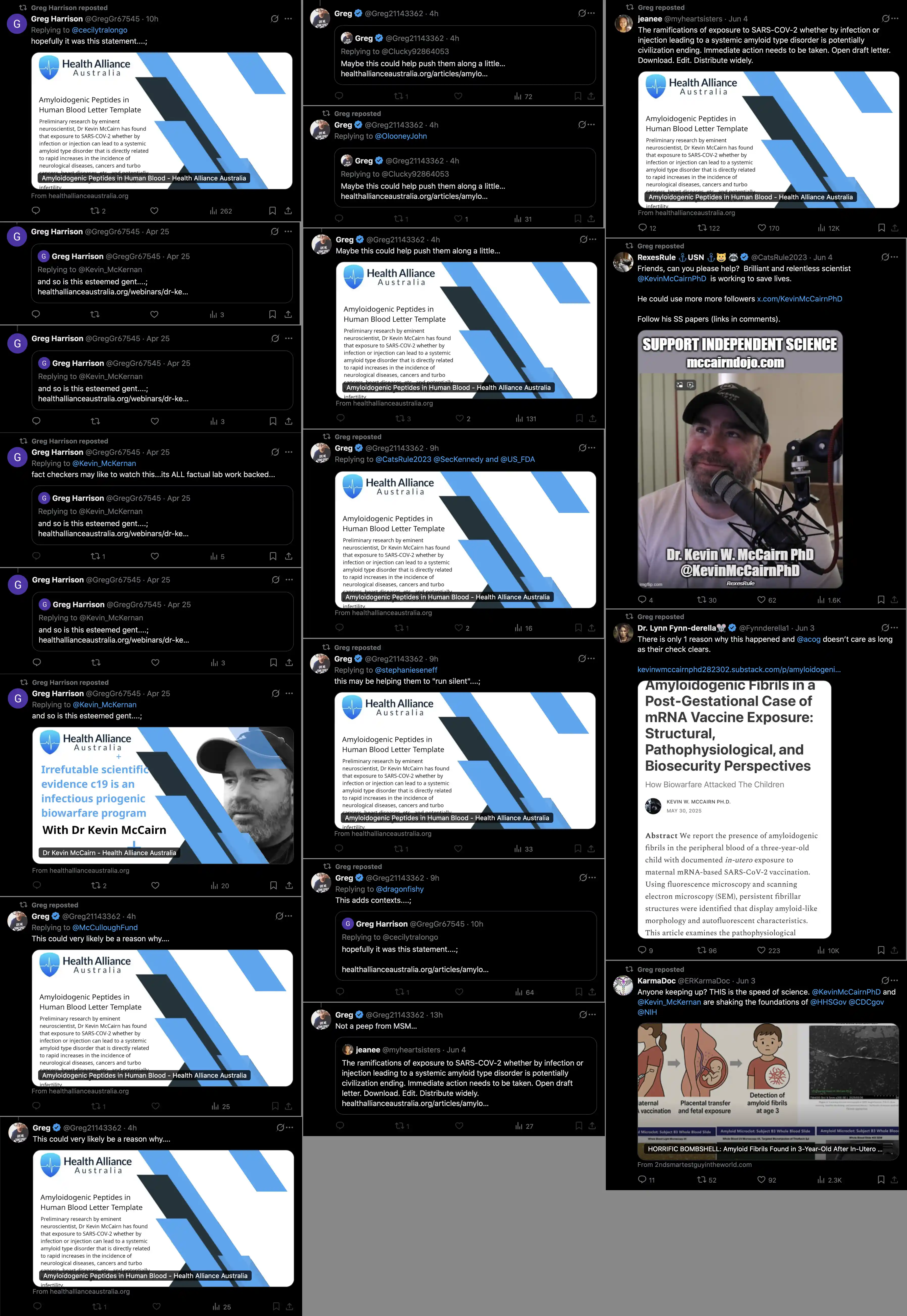
In March 2025 McCairn did an experiment where he injected pieces of Hirschman's clot into the heart of a hamster, which died right after the injection. He didn't do a public stream about the experiment, but he only livestreamed it on his Discord.
Wayne Crouch made an infographic about how McCairn killed the hamster
after injecting it with "our amyloid jab clot
samples". Crouch said that McCairn was linked to Solution of
Scientists, which is the group that Crouch and Greg Harrison are part
of, and which I'm calling the "Aussie Quinta
Columna". Crouch is supposed to be a journalist, but in his
infographic he somehow managed to misspell the words "scientist's", "hampsters", and "acess": [https://

Wayne Crouch also wrote a Facebook post where he referred to the
people who conducted McCairn's experiment as "we": [https://

Crouch claimed that McCairn's experiment was proof that "we will see the rapid death to the shot recipients". But if the hamster died within seconds from the injection, then why haven't vaccinated people died rapidly yet, even though years have already passed from vaccination? And anyway, if McCairn injected protein into the heart of the hamster, it's hardly comparable to injecting LNPs filled with RNA into a muscle in the arm.
In March 2025, Tom Haviland wrote a Substack post where he described
McCairn's analysis of the clots. [https://

Haviland's Substack post had a document attached named
Kevin_, which described the results of
McCairn's analysis of the clots. The authors of the document were
anonymous, and the document was mysteriously signed by "The Researchers":

McCairn told me he was not involved in writing the document. However
one of the authors was probably Greg Harrison, because the document was
formatted in a similar way as Greg's documents. In 2024 Laura Kasner
posted another similar PDF about the clots, where the PDF said that the
"authors remain anonymous for safety
reasons", but Kasner wrote that Greg was one of the authors.
[https://
The document about McCairn's findings said that "ORF-19 and ORF-11 are now functionally implicated in the induction of prionic seeding":

The names of ORFs were written with a hyphen in the document. The
names of ORFs are normally written without a hyphen, but Greg Harrison
and Wayne Crouch have both written the names of ORFs with a hyphen.
[https://
McCairn told me on his Discord: "You seem
incapable of understanding that I am working independently and the only
link is that is that Greg claims to have done some investigation on the
clots. What he has done has nothing to do with my lab analysis."
But I linked to the Kevin_ document and
I asked him: "If you're so independent from Greg et
al. then why did they write this report about your findings?" And
McCairn replied: "I have no idea, and I did not give
them permission to write that report or associate it with me any
way."
Kevin McCairn's website has a page called "Prion
Research Investigation Project", which includes a stock photo of
people in a lab that I thought looked similar to the stock footage in An
Unholy Triad: [https://

But then I found that McCairn's stock photo came from the same
collection of stock footage that was used in An Unholy Triad: [https://


I thought that the stock photo on McCairn's website might be a clue that the same people who made An Unholy Triad were also involved in making McCairn's website. McCairn told me that the page on his website with the stock photo was made by Chris France, who has also made other parts of McCairn's website. But when I asked Chris France how he picked the stock image, he didn't answer me. So the question of whether the matching stock footage was a coincidence or not is still open.
Greg's AI seems to have prophesied the results of McCairn's Rt-QuIC
analysis, because in December 2024 Greg wrote: "Thx
Kevin, will send you emails with word docs attached in which AI is now
telling us we have identified a new blood-born Amyloid-prion disease.
All is conjecture but AI seems highly convinced we shall soon identify
this new Amyloid-Prion hybrid disease with deep NMR's & RT-QuIC,
plus a few more techniques to properly nail it. Interesting times
ahead..thx Gregh" [https://
Greg Harrison, Wayne Crouch, and Lisa Johnston are all promoting a
McCairn-style narrative about a coming prion apocalypse. Lisa Johnston
tweeted that "Planet earth is under attack by
prionic amaloidosis created by man": [https://

Wayne Crouch was credited as the director of the Unholy Triad videos.
The final video in the series presented a scenario of a prion
apocalypse. First vaccinated people started developing calamari clots,
and then prions from the clots ended up in wastewater, and wastewater
carried the prions into the sea. Then the seawater evaporated and formed
into deadly prion rain, and also the prions spread to humans who ate
fish, and in the end all of humanity died: [https://

In February 2025 Kevin McCairn appeared on a video panel about the
clots hosted by Steve Kirsch, which also featured Kevin McKernan,
Richard Hirschman, Tom Haviland, and Greg Harrison: [https://

Kirsch is connected to the story about the clots in multiple ways,
because I believe he was the second person after Jane Ruby who
interviewed Hirschman, and the first person who interviewed Anna Foster
and Cary Watkins, who to my knowledge were the next two people after
Hirschman who said they had seen the clots. Jane Ruby wrote that Kirsch
worked with Epoch Times to put out his own version of the story about
the clots, but they left her evidence out of the story. [https://
So I thought that McCairn might have somehow become involved with the
clots through Kirsch, because Couey said: "Steve
Kirsch wanted me to evaluate Kevin McCairn's grant proposal in 2022, and
we had a nice little text exchange on my phone about that. And so there
again, he even offered to send me to Tokyo to help him work, and help
him do those experiments." [https://
So perhaps it was a coincidence that McCairn happened to send the grant proposal to Kirsch, because McCairn doesn't seem to otherwise be too closely connected to Kirsch.
In March 2025 when I compiled evidence on McCairn's Discord that
Greg's ORFs were a hoax, McCairn recognized the ORFs to be fake. Then in
April 2025 Greg Harrison tweeted: "And at this point
in time, wish to retract my ORF 'rantings'
due to our lack of credible evidence that any of our ORF stuff actually
exists". [https://
When I asked McCairn on his Discord how he first got connected to Greg, he replied: "Here @Henjin is a screenshot of first emails, with time stamps with respect to Greg and Richard. Why don't we start here so you can order your thinking a little better. As memory serves I was involved in a Skype call, it was a group of researchers who had been looking at histological sections of blood from patients that showed microclotting where they presented their histological findings and I initially advised them on how to proceed to stain their tissue for presence of amyloid." And later McCairn also explained: "As I recall it was meeting with clinicians who have worked with Richard to begin to measure and quantify what the clots were. They were trying to do thioflavin staining, but lacked the equipment necessary for precise histology. He [Greg] was a part of that call, I didn't know him but he was obviously co-ordinating with Richard and the clinician group in Alabhama who are treating amyloidogenic microclots. As they were making procedural errors in trying to type the tissue, and I had the facilities available, i offered to process the tissue properly so that histological staining for amyloids was done correctly. I have received no money from them to do this or for the subsequent anlayses I have done using RT-QuIC, SEM/EDX and Raman spectroscopy. All of those methods have confirmed an amyloidogenic signature. Does that make sense?"
So as of now it's more or less clear that Greg's crew is deliberately producing disinformation, but it's not yet clear if McCairn is complicit in their operation, or if McCairn rather got inadvertently involved with Greg's crew because he volunteered to help with the histological staining analysis, or because he was looking for samples of the clots to analyze.
But in the case that the clots are fake, and Harrison and Hirschman
know themselves the clots are fake, then would they trust someone to
analyze the clots who is not in on the scam? People who had presented a
laboratory analysis of the clots before McCairn include Mike Adams, Ryan
Cole, Ana Mihalcea, Clifford Carnicom, Diana Wojtkowiak, Zandre Botha,
Arne Burkhardt, and Greg Harrison. But I believe all of them are
controlled opposition. (I explained why I think
Burkhardt and Cole are controlled opposition on my page about turbo
cancer, because the term "turbo cancer" was
introduced in Burkhardt's conference, and I believe Cole was the first
major person who promoted the hoax about turbo cancer but who was not
German: turbo.
In March 2025, Philip McMillan did one video about McCairn's analysis of the calamari clots, one video where he talked about Greg's ORFs and how McCairn had found that the clots were autofluorescent, and another video about an AI-generated document that had mystery ORFs going up to ORF100:

When I asked Grok to list people who have presented a laboratory
analysis of the calamari clots, the first two people it listed were Mike
Adams and Greg Harrison. Grok said that McCairn's Raman and RT-QuIC
analysis was done by "Harrison's team",
because Grok cited a Substack post by Nicolas Hulscher, who gave the
impression that McCairn's analysis was done by Greg's team: [https://

Added in June 2025: Over a period of only two days, Greg Harrison
posted all of these tweets that promoted McCairn: [https://
Even after I exposed Greg's ORF hoax, McCairn only threw grew halfway under the bus, because McCairn acted like Greg was just some foolish boomer who got tricked into believing in his own AI hallucinations (even though in reality Greg was clearly part of a coordinated disinformation operation that also included Wayne Crouch and Lisa Johnston):
I think McCairn himself represents another arm of the disinformation operation, so even though he distanced himself from the Solution of Scientists crew, he hasn't drawn attention to the ORF hoax as evidence that the clots are not real. And when the SOS crew released the trilogy of preprints about the clots in 2026, McCairn failed to mention how me and a moderator on his Discord had earlier shown how the data in the trilogy was fake. And he hasn't drawn attention to how Hirschman and Haviland have continued to support Greg even after Greg was shown to have fabricated the data about the ORFs.
In early 2025 when Greg Harrison was promoting the hoax about the
fake ORFs, his biggest cheerleaders on Twitter were users called
CoyoteSanctuary and RexesRule/CatsRule2023. Both of them also joined
Kevin McCairn's Discord around December 2024, which was after McCairn
had started doing videos with Hirschman and Harrison. Even after I had
posted exhaustive evidence on the Discord that the ORFs were fake,
RexesRule and CoyoteSanctuary kept defending Greg's ORFs, and they just
told me that I was crazy or that I was a counterintelligence agent. So I
thought they might have been in on the scam, since otherwise their
behavior of defending Greg's hoax did not seem reasonable: [https://


When I looked into the Twitter profile of RexesRule, I noticed that
her banner image had a seal that said "NAVSECGRUDIV" and "NAVCAMSEASTPAC": [https://

Wikipedia says: [https://
The Naval Security Group (NAVSECGRU) was an organization within the United States Navy, tasked with intelligence gathering and denial of intelligence to adversaries. A large part of this is signals intelligence gathering, cryptology and information assurance. The NAVSECGRU organization was active from March 1935 to September 2005.
In addition to being part of the Navy, NAVSECGRU was also part of the National Security Agency's Central Security Service.
The NAVSECGRU organization was transferred to the Naval Network Warfare Command (NETWARCOM) where its former assets made up the Information Operations Directorate.
"NAVCAMS EASTPAC" is short for "Naval Communication Area Master Station, Eastern
Pacific". [https://
I found a tweet where RexesRule wrote: "My
NAVSECGRU team was the best." [https://

An announcement from 2005 said: "What was
formerly NAVSECGRU has now become NETWARCOM's Information Operations
Directorate." [https://
Later after I got Kevin McCairn to agree with me that Greg's ORFs
were fake, Greg posted a tweet where he wrote "And
at this point in time, wish to retract my ORF 'rantings' due to our lack of credible evidence
that any of our ORF stuff actually exists." [https://
But on the other hand it's possible that RexesRule was not part of
Greg's disinformation team in the way that Wayne Crouch seems to be part
of a coordinated team with Greg, because the oldest tweet that matched
the query @ was only posted in
February 2025. And similarly the oldest tweet that matched the query
@ was only posted in December
2024. But before that both RexesRule and CoyoteSanctuary had frequently
interacted with Kevin McCairn. So RexesRule seems more like a
cheerleader for McCairn, who started promoting Greg's ORF hoax after
Greg had appeared as a guest on McCairn's stream.
I think CoyoteSanctuary is likely to be a useful idiot, but I'm not so sure about RexesRule, especially considering how McCairn is also connected to the US Navy or US naval intelligence through Charles Rixey (who is a former Marine, and who received the DEFUSE proposal from Joseph Murphy who works for the Office of Naval Research), Spartacus (who is a merchant marine), Paul Cottrell (who had a radio show on a network operated by a naval intelligence agent), J.J. Couey (whose former boss works for the ONR), and Addy Adds (who lived on US naval bases, and who ghostwrote a book for a Qtard lady whose channel was managed by someone who also managed the channel of an ex-Marine Qtard).
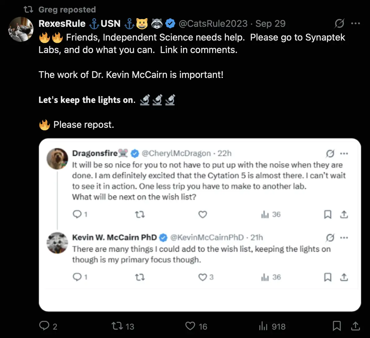
RexesRule has made many meme-style images where she has promoted Kevin McCairn, and she frequently posts tweets where she asks people to donate money to McCairn:


Greg Harrison also retweeted a tweet where RexesRule asked people to
donate money to McCairn: [https://
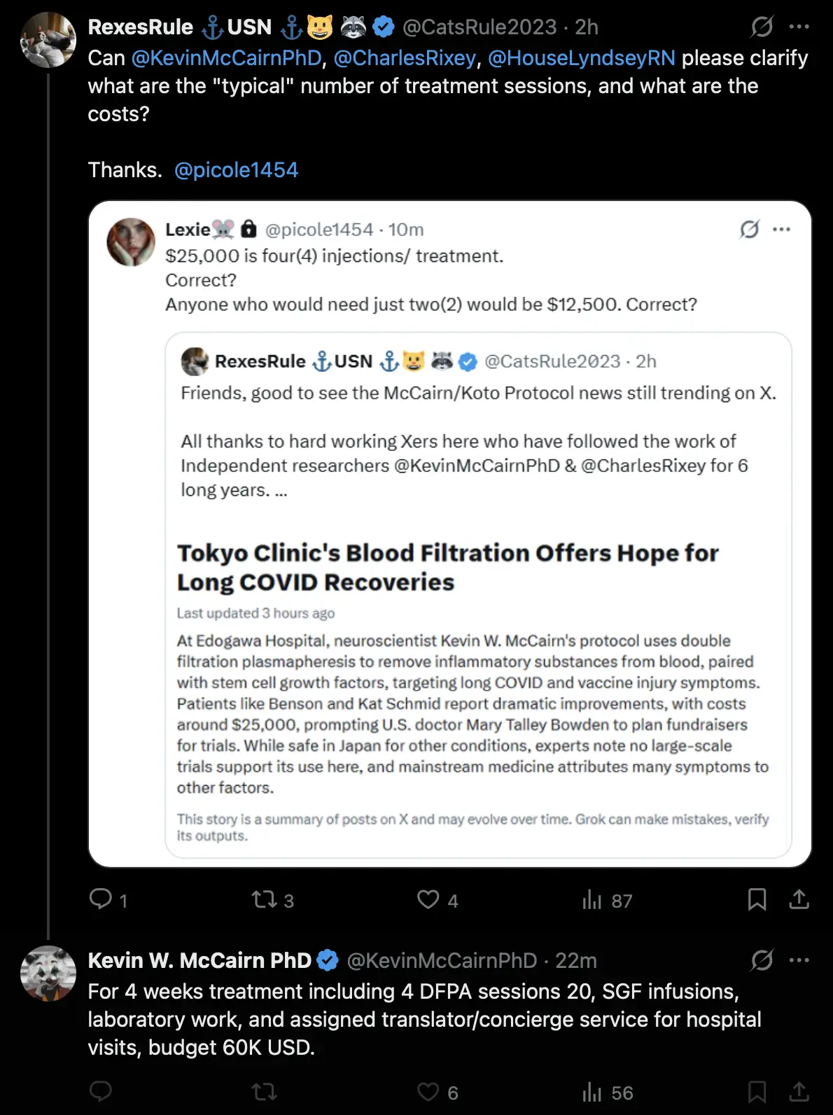
Added in 2026: RexesRule has now started making infomercial-style
memes to promote McCairn's amyloid fibril removal scam: [https://

Now you too can get your fibrils removed for the low-low price of
60,000 US dollars: [https://
Added in June 2026: RexesRule has now started making videos promoting
McCairn, because she says she is trying to get McCairn's work noticed by
the mainstream media: [https://

In July 2026 RexesRule promoted this video of Greg Harrison, even
though I had shown her many times that Greg had fabricated his data:
[https://


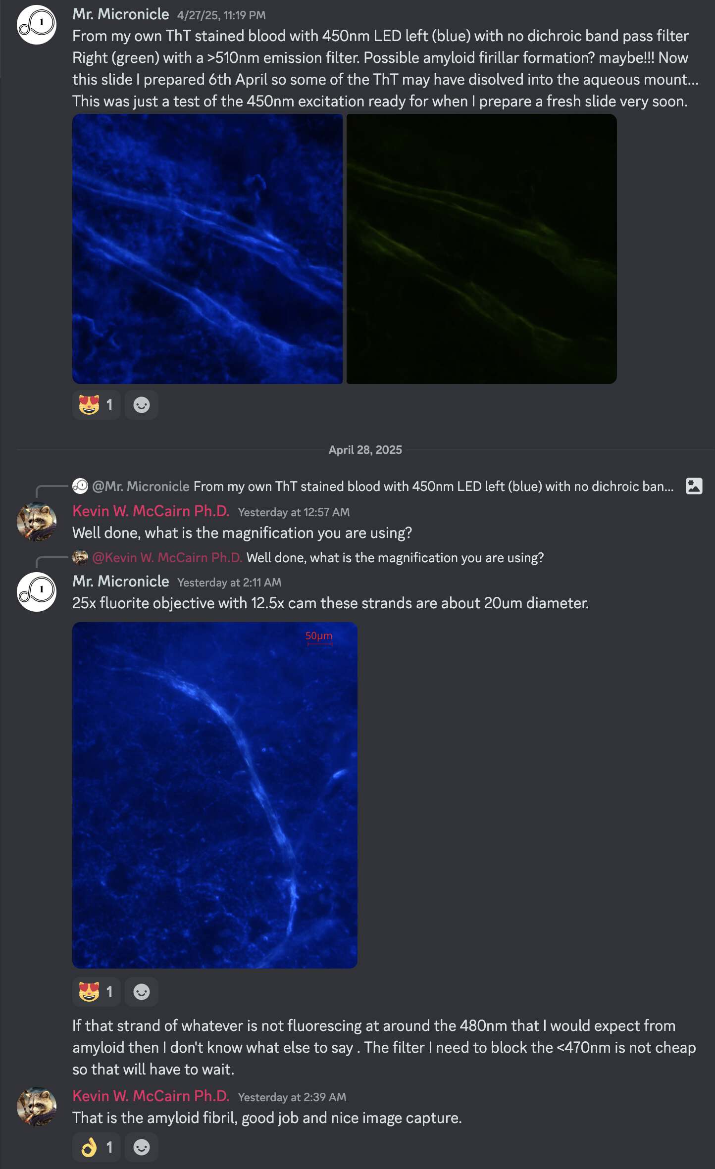
A user called Mr. Micronicle joined Kevin McCairn's Discord server in 2025. He posted various microscope images on the server, including images that he claimed showed calamari clots that came from the body of a dead person, even though he refused to answer me where he obtained the clots.
Mr. Micronicle said that he took microscope photos of his blood stained with Thioflavin T, and he showed an image of a fiber-like object in the blood with a diameter of about 10-20 µm. Then Kevin McCairn said "That is the amyloid fibril, good job and nice image capture":
I told McCairn:
Wouldn't it be an aggregate structure that consists of multiple fibrils? A single fibril would be much narrower based on what ChatGPT said:
Amyloid fibrils are very thin protein aggregates.
Their diameter typically ranges from 7 to 13 nanometers (nm), regardless of where they form (blood, organs, brain, etc.).
In blood-related diseases like AA amyloidosis or AL amyloidosis, amyloid fibrils circulate or deposit, and their size remains in that typical range.
Cryo-EM (cryo-electron microscopy) and atomic force microscopy studies have shown that some fibrils can be as thin as 6 nm or up to about 15 nm, depending on the specific protein involved and conditions of formation.
McCairn responded: "All peptides are aggregate structures. Depends on the method of aggregation though and their resistance to proteases as to how we classify them as pathogenic." But I showed that ChatGPT said:
In scientific terminology:
A fibril is understood to be a very thin, elongated structure, usually in the nanometer scale for diameter (approximately 7-13 nanometers for amyloid fibrils).
Structures that are micrometers wide (especially 10 micrometers, which is about 1,000 times thicker than a typical fibril) would more accurately be called:
Bundles of fibrils
Aggregates
Fibers or fiber-like structures (sometimes used when many fibrils associate)
Thus, a 10 micrometer-wide formation would be considered a fiber or an aggregate of fibrils, rather than a single fibril itself.
And McCairn said: "It is a fibril, singular, of oligomerized amyloidogenic fibrin. That description is correct." But I showed him that ChatGPT responded:
In principle, no - if you are using a standard light microscope, it would not be possible to image a single fibril of oligomerized amyloidogenic fibrin.
Here is a detailed explanation:
A single fibril (even one formed by fibrin adopting amyloid-like structure) would have a diameter of around 6-15 nanometers, sometimes slightly thicker depending on conditions.
The diffraction limit of optical microscopy (~200-250 nm) means anything thinner than that cannot be resolved as a distinct object.
Therefore, under a standard light microscope, what you would see would be aggregates, bundles, or dense regions of multiple fibrils - not an isolated, singular fibril.
Wikipedia says: "Fibrils (from Latin fibra) are structural
biological materials found in nearly all living organisms. Not to be
confused with fibers or filaments, fibrils tend to have diameters
ranging from 10 to 100 nanometers (whereas fibers
are micro to milli-scale structures and filaments have diameters
approximately 10-50 nanometers in size)." [https://
McCairn's defense was that the calamari clots were a novel phenomenon that required a novel vernacular, so it was acceptable for him to use the word "fibril" in an unconventional sense. He told me: "Trying to use old vernacular to a new blood prion disorder will lead the spergs like you to blow a fuse, but you'll just have to put up with it."
Then McCairn said this about the clots: "Nothing @henjin is sore that there have been multiple replications of the same phenomena and that they satisfy the metrics required to call them amyloidogenic peptides." But I replied: "You mean the fibrin or other proteins in the clots contain segments that act as amyloidogenic peptides? It wouldn't make sense to say that the clots themselves are amyloidogenic peptides." And I pointed out that ChatGPT said:
Strictly speaking, no, it is not accurate to say that fibrin clots themselves are amyloidogenic peptides. Here is the proper interpretation:
A peptide refers to a small chain of amino acids, typically much shorter than full proteins.
Fibrin is a full-length, large polymeric protein (~340 kDa before cleavage), not a small peptide.
A fibrin clot is a macroscopic network formed by polymerized fibrin monomers, often extending hundreds of micrometers across - not a single molecular entity or a simple peptide.
Thus:
A fibrin clot is not itself a peptide. It is a supramolecular structure made of many fibrin molecules.
Amyloidogenic peptides are smaller fragments or sequences capable of forming amyloid fibrils on their own.
In pathological conditions, portions of the fibrin molecule become amyloidogenic - but the entire clot should be referred to as an amyloid-rich structure or amyloid-like fibrin network, not as a "peptide."
Accurate phrasing would be:
"Fibrin clots can adopt amyloid-like structures."
"Amyloid fibrin contains amyloidogenic regions within the fibrin protein."
"Pathological fibrin clots exhibit amyloid features."
Then McCairn just said that "we are dealing with a novel phenomenon, and as such the current repertoire of nomenclature does not fit well". But he wasn't willing to admit that he used the wrong terminology.
Earlier I had also called him out for saying that fibrin was a peptide, even though a peptide is a short chain of about 2-50 amino acids, but fibrin is a polymerized protein product of fibrinogen, and each fibrinogen unit in turn is made up of 6 proteins that each have a length of about 400-600 amino acids. McCairn's response was that "You should also be reminded that the vernacular peptide and protein are interchangeable." But I pointed out that ChatGPT said:
No, it would not really be correct - even in casual speech - to call fibrin a peptide.
Here's why:
- Fibrin is very large - it's made by polymerizing fibrinogen, which itself is almost 3000 amino acids long.
- Even a single fibrin monomer (before polymerization) is way too big to be called a "peptide."
- Peptides usually refer to short chains - dozens, or maybe a few hundred amino acids at most.
Calling fibrin a peptide would sound wrong to most people familiar with biology, even casually. It's much better (and more natural) to call fibrin a:
- Protein, or
- Polymerized protein, or
- Fibrillar protein, depending on context.
If McCairn sees a random fiber under a microscope, he says the fiber is an amyloid fibril, in the same way that Ana Mihalcea would say that the same fiber is a Morgellons filament or a carbon nanotube, and Mike Adams would say that the fiber is a reptilian nanowire interface structure.
Mr. Micronicle's website used to be called "Zero
Infinity One Network", which he said was "a
reference to the esoteric sense of what mount Zion represents in
esoteric terms": [https://

His Telegram channel was called "ZIO.NETWORK". [https://
Added in 2026: Greg's AI now told him that "fibril-like peptides" had been found in the
laboratory, so the AI managed to combine McCairn's incorrect uses of
both the words "fibril" and "peptide" into a single term: [https://
In the thread below, Richard Hirschman quoted a tweet by McCairn's
naval intelligence cheerleader RexesRule, who posted McCairn's SEM image
of one of Hirschman's clots. Hirschman said the image showed amyloid
fibrin, but a biologist called Ian Musgrave said it was not correct:
[https://
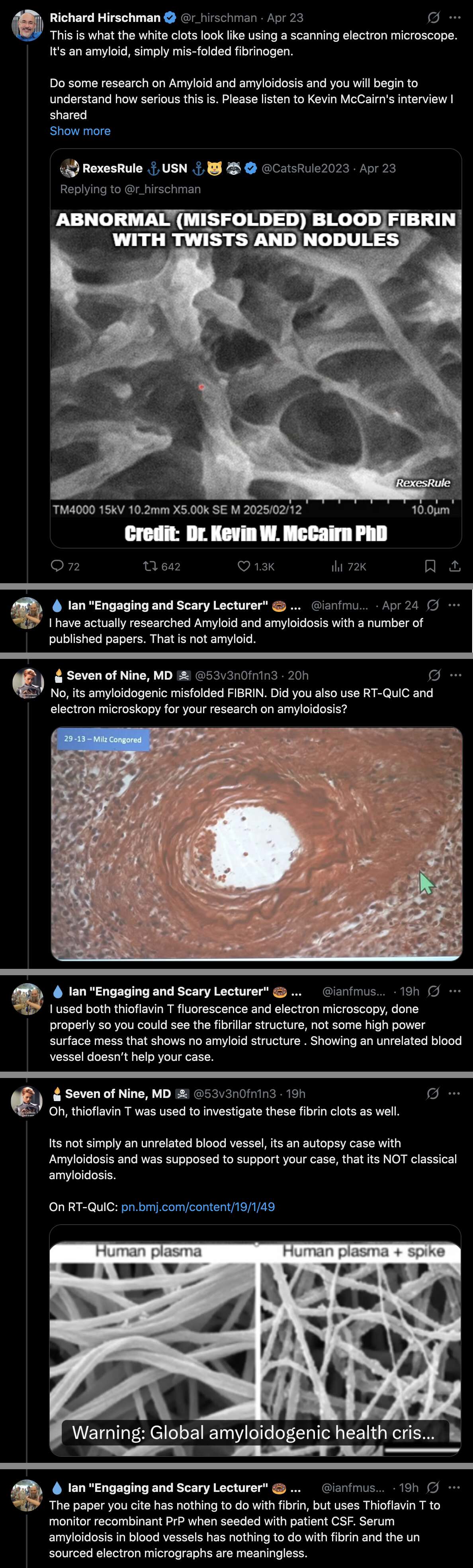
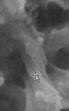
In a stream McCairn did about the SEM images, he showed this image:
And he said: "When you go down to 5,000 times,
what you see here - and it's only because unfamiliar with looking at
these structures, that this was a standout abnormal to me, right - there
are very clear nodular forms on these peptides, ok. I don't know if you
can see that, right, so my cursor is here, right, like this will stick
out. And if we look at this primary branch here, what you see is -
fibrin should be a long smooth rope-like peptide that essentially just
overlays itself to form the network around which platelets and other
tissues that form a clot - can aggregate, ok. Here what you're looking
at is a - and so, you're looking at very abnormal structural properties
that are standout to someone who's familiar with looking at peptides in
and of themselves, ok. And those are these nodular forms, and also -
what I would point out to people is that - pay attention to this thicker
filament, right, and um, in biology, often what you see - and that's not
the best example that I can give for people to think about - is often
when you look at a tree, a tree, as you look at the trunk has a sort of
twisting effect to it, right. It sort of starts at the roots, and it
sort of has a rotation to it, as it goes up to form the branch area of
the tree. But the trunk often, if you pay attention to it, you'll see it
has a rotation to it. And generally in biology, I would say it's a
right-hand rotation, it's the right hand rule of thumb, and you can get
into all sorts of metaphysics around electrodynamics and what that - how
that relates to the body. But in this instance, what you're seeing is
that you're seeing a faster twist - so rotations per unit of distance -
than you would expect to see in normal healthy tissue." [https://
He kept saying that fibrin was a peptide, even though fibrin is not a peptide but a protein. Peptides are short chains of about 50 or fewer amino acids.
The reason why he said that a twisted shape was somehow characteristic of misfolded fibrin might be because earlier he claimed a similar shape was characteristic of microclots made of misfolded fibrin, even though the twisted fibers he showed under a microscope were likely not even made of fibrin, but they were probably textile fibers or cellulose fibers that he misidentified as fibrin. But the so-called "primary branch" in his image has an irregular shape which looks like it might possibly be due to similar lengthwise twists, even though it's not clear if that's the case or not:
The fibers McCairn showed earlier were about 10-30 µm wide, but his so-called "primary" branch is only about 2 µm wide. McCairn has not presented good evidence that a twisted shape would be a characteristic of structures made out of misfolded fibrin, or that the twisted shape would occur in different types of structures with diameters at different orders of magnitude.
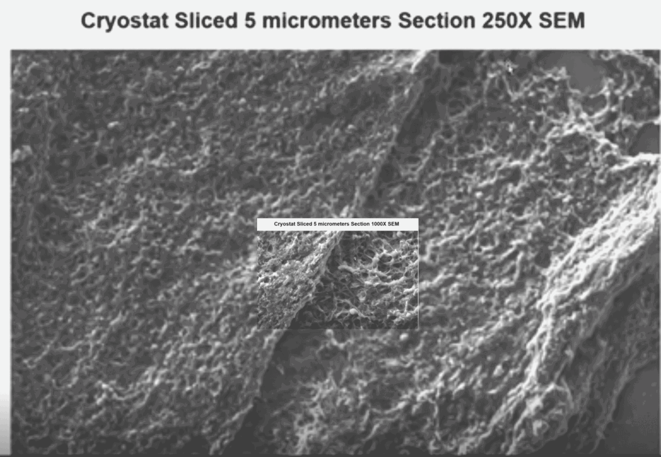
McCairn also showed the images below of the same sample at 250x and
1000x magnification (edited later to take
higher-quality images from McCairn's Substack post): [https://
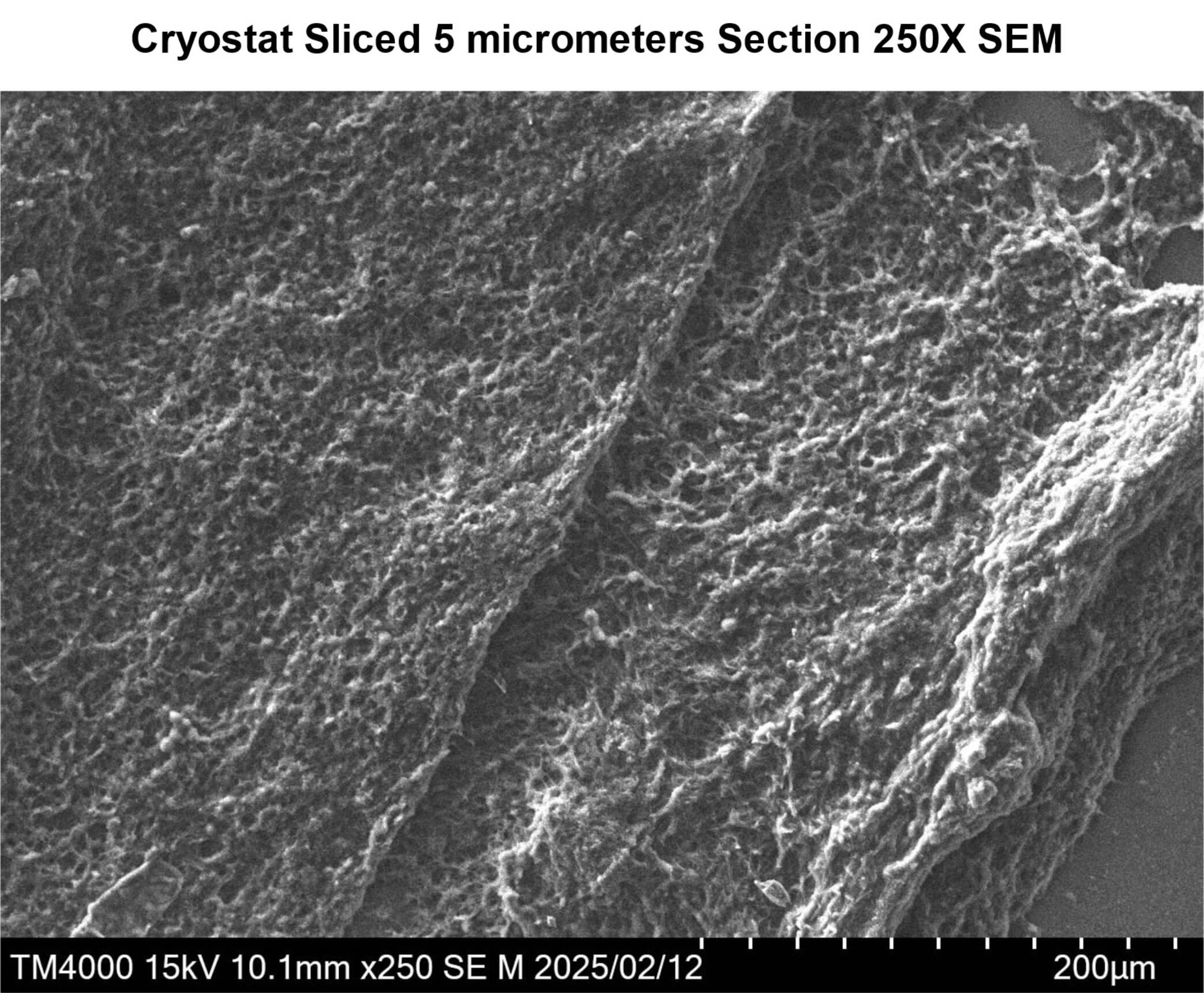
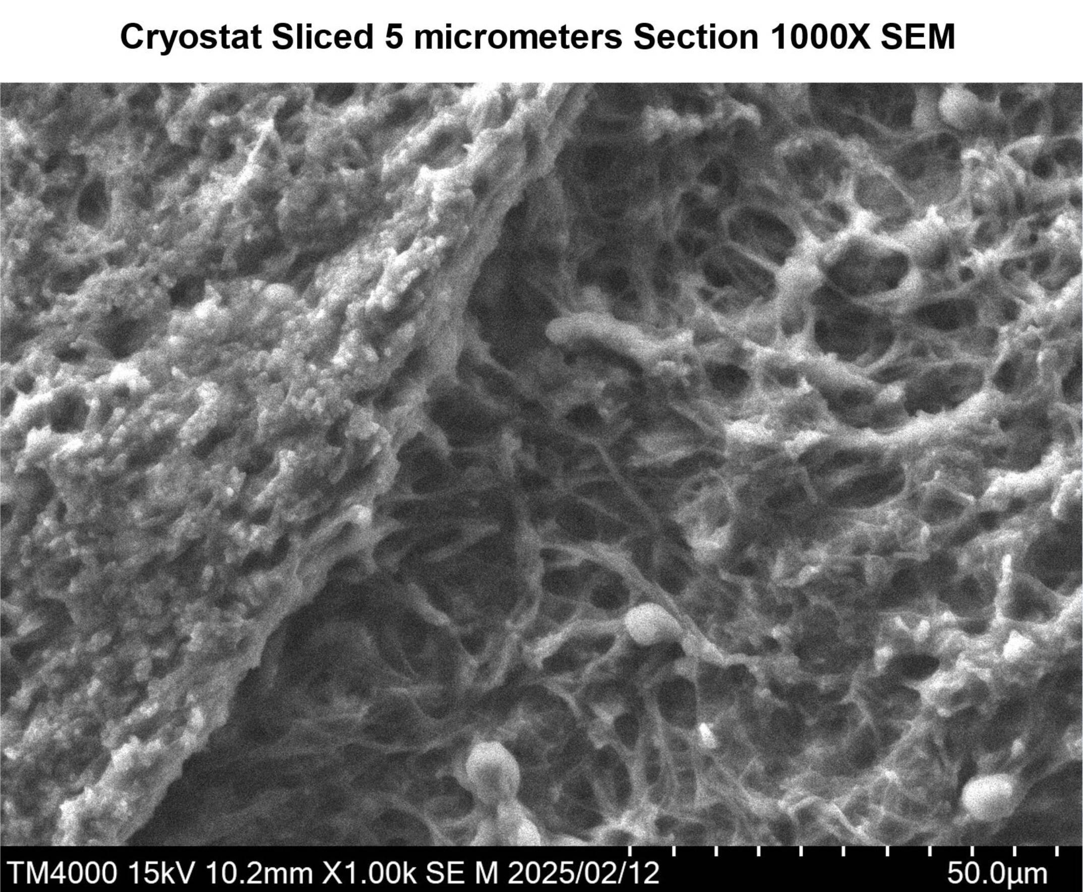
The GIF files below show the 1000x image overlaid with the 250x image, and the 5000x image overlaid with the 1000x image. The 5000x image doesn't have a much higher level of detail than the 1000x image:

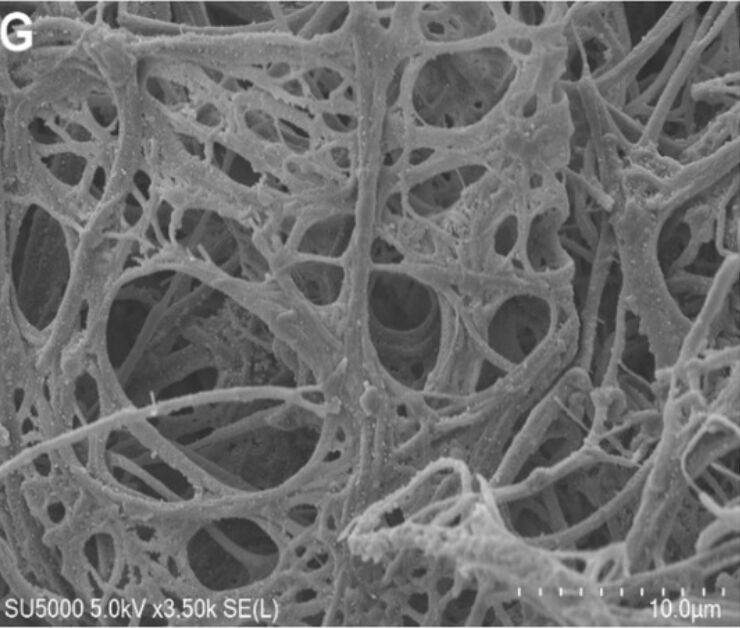
Below McCairn's 5000x image is shown next to an SEM image of a
regular fibrin clot at 5000x magnification. [https://

A typical diameter for a fiber of fibrin is about 0.1 µm: [https://

When I tried to find SEM images of fibrin clots that resembled
McCairn's image, so that had they wide branches with an irregular
structure, I found the image below. Image A shows regular spaghetti-like
strands of fibrin that have a fairly uniform diameter of about 0.1 µm.
But in image B where fibrin was clotted in the presence of the lipid
DPPC, the fibrin formed wide irregularly shaped structures, so some
branches of the structure have a diameter above 1 µm like in McCairn's
image. Image B was described like this: "In
contrast, in the DPPC MLV suspension the heterogeneous large-pore gels
are formed with thin branched fibers and lipid particles incorporated
into the gel structure (Figure 1, B). The
images reveal many free fiber terminations. Fibers appeared to be highly
adhesive forming dense mats and tight bundles." DPPC is a lipid,
and MLV means multilamellar vesicle which is a type of liposome, so the
balls circled in image B are balls of lipid: [https://

I also found a paper where fibrin clots were created in the presence
of a fibrinolytic compound, which resulted in the formation of wide
strands with a diameter of about 0.5 to 3 µm: [https://
The branches in McCairn's image had a diameter of about 0.2 to 3 µm. ChatGPT said that fibrin structures with a diameter on the micrometer level are unlikely to consist of individual fibrin fibers, but rather bundles of multiple fibrin fibers glued together:
Under normal physiological conditions, fibrin fibers (the building blocks of a blood clot) have diameters typically in the range of 50 to 200 nanometers (nm), sometimes up to 500 nm. So 0.05-0.5 µm is usual.
However, under abnormal or pathological conditions, fibrin fibers can become thicker, but usually not to the extent of 1 µm, and very rarely close to 10 µm as single fibers.
[...]
Bundles of fibers can easily be 1-10 µm in diameter - but those are multiple fibers glued together, not single fibrin polymers.
Single fibrin fibers, even under weird conditions, very rarely cross the 1 µm diameter threshold.
However later ChatGPT said that even fibers of fibrin with a diameter over 1 µm might not necessarily consist of multiple narrower fibers glued together, but wide fibers might also be formed due to a greater degree of lateral aggregation in the stage where the protofibrils form into fibrils:
Thick fibrin fibers with diameters over 1 micrometer generally form through a similar basic polymerization process as regular thin fibers (around 0.1 micrometer), but there are important differences in how they become thick.
Both thick and thin fibrin fibers start with the same initial steps: thrombin cleaves fibrinogen to create fibrin monomers, which then align end-to-end to form protofibrils, and protofibrils laterally aggregate to form fibrils. In thinner fibers, this lateral aggregation is moderate - a few protofibrils come together to form a fibril, and those fibrils form relatively fine fibers.
For thicker fibers, the key difference is that more extensive lateral association occurs. Multiple protofibrils aggregate side-by-side more completely, and sometimes additional bundling happens between already-formed fibrils. There is evidence that under certain conditions - such as low fibrinogen concentration, low ionic strength, or altered thrombin activity - multiple narrow fibers can clump together after initial formation, effectively fusing into wider fibers.
Thus, thick fibers (>1 µm diameter) can result from both mechanisms:
More extensive lateral aggregation during initial assembly, leading to inherently thicker fibers.
Post-assembly bundling or merging of multiple narrower fibers.
If indeed McCairn's image does even show a clot that is made of fibrin, then I don't know if the thickest branches in his image are bundles of multiple strands of fibrin joined together, or if they are just single wide fibers of fibrin. McCairn said that his image showed misfolded fibrin because the branches had an irregular and twisted shape, and the branches didn't look like smooth spaghetti like regular fibrin. But maybe the irregular shape was if the branches were made up of bundles of fibrin and not individual strands of fibrin. (And the clumping of the strands may have been if for example the fibrin structure formed in the presence of a lipid or a fibrinolytic compound, and not necessarily because the fibrin was misfolded.)
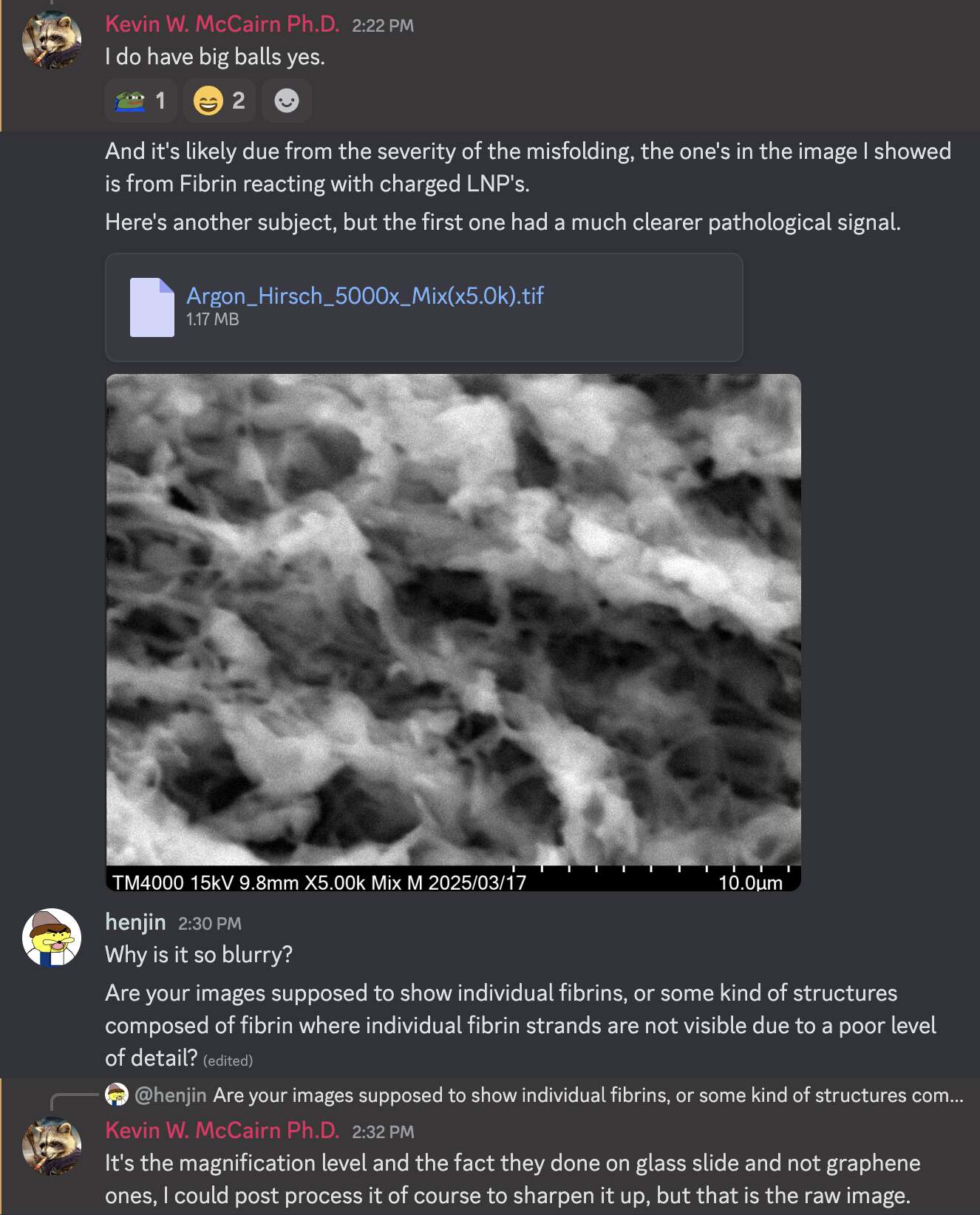
When I asked McCairn on Discord why his SEM image had such a poor level of detail, he said: "The difference in detail comes from using a graphene base vs glass slide, and other factors, backscatter acquisition from surface or combined." When I asked why he couldn't use a graphene base, he said: "I use glass to be able run Raman on the same sample."
He also posted another similarly blurry SEM image at a 5000-fold magnification level (but it does seem to be a real SEM image judging from the EXIF metadata, which even includes the serial number of the Hitachi TM4000 instrument):
I initially questioned if McCairn's images were really even taken
with an SEM, because McCairn made it seem like he had bought his own SEM
instrument, which would've seemed like an unnecessarily expensive
purchase, but I didn't see an SEM in his lab in his videos, so I
questioned if he had actually bought an SEM. For example in a video
where he showed his SEM images, he showed a photo of the Hitachi TM4000
SEM, and he said: "It costs serious money to go and
get this type of data, ok. The machine you're looking at there is
hundreds of thousands of dollars." [https://
But McCairn told me on his Discord that he didn't end up buying his
own SEM instrument, but he visited a lab in Japan to do the SEM and
Raman analysis, and he linked to an old video where he visited the same
lab and used the Hitachi TM4000 microscope. [https://

Hirschman also said: "We've got now Kevin McCairn
who's done this work. It costs lots of money. This equipment that he
uses costs hundreds of thousands of dollars. It's not like you can just
walk up somewhere and have this work done." [https://
One of McCairn's followers went around asking billionaires to donate
money to McCairn, because he said McCairn "LITERALLY
JUST NEEDS A FEW HUNDRED THOUSAND DOLLARS IN EQUIPMENT AND COULD SAVE
EVERYONE FROM DYING HORRIBLY": [https://


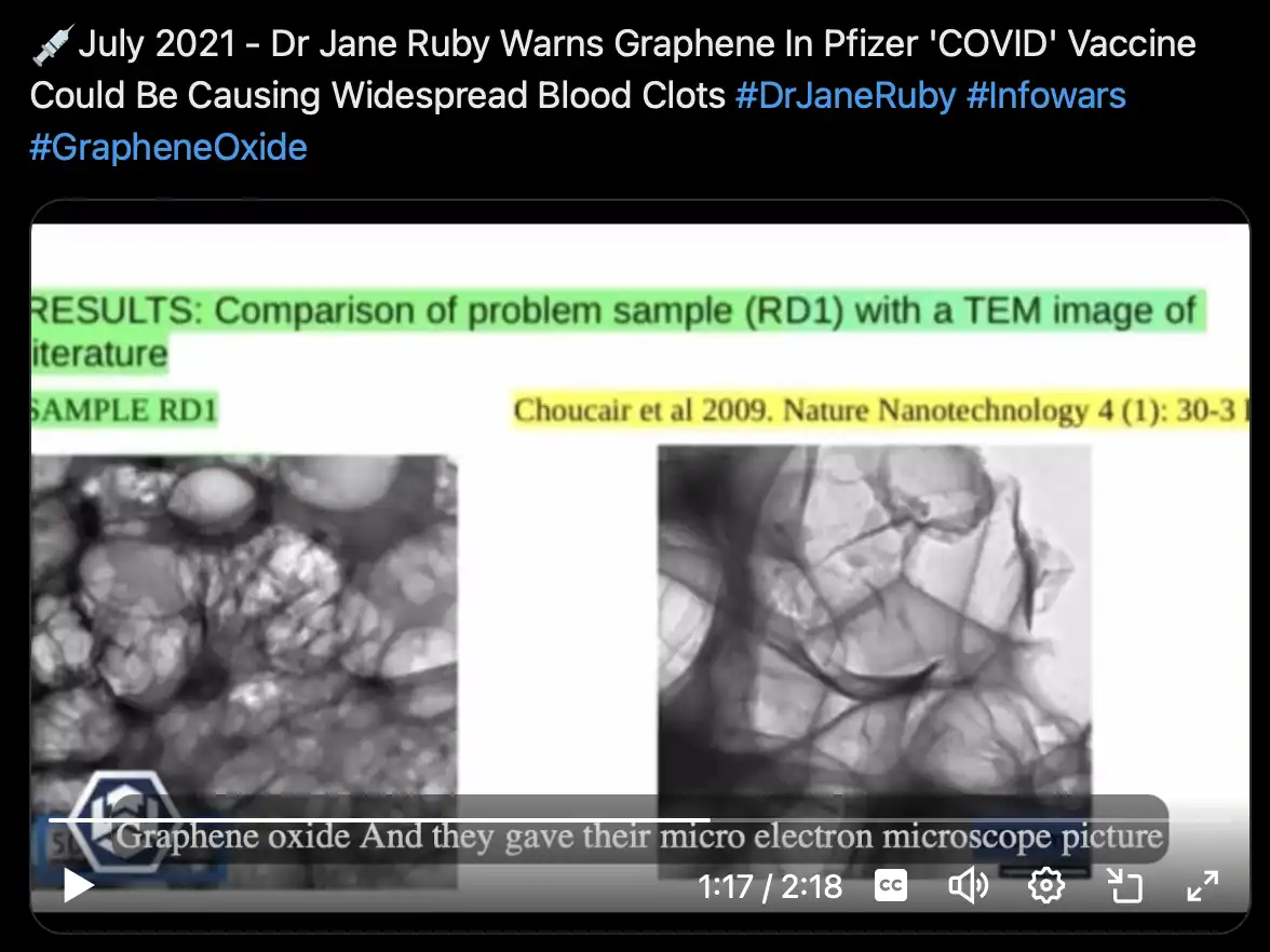
In 2021 Jane Ruby said that an electron microscope image of COVID
vaccines looked similar to graphene oxide, which she presented as
evidence that COVID vaccines contained graphene oxide. It's reminiscent
of McCairn's operation of taking SEM images of the clots: [https://
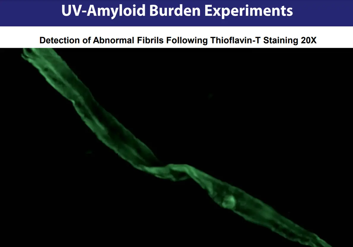
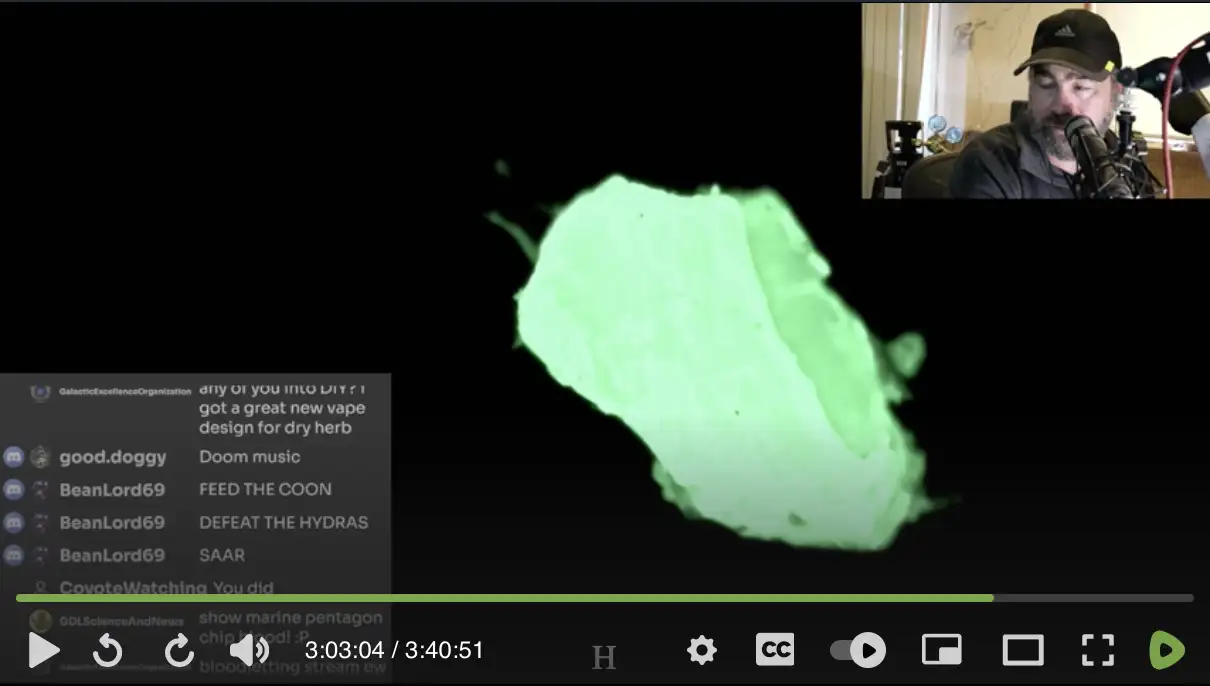
McCairn claimed that the image below showed a fibril he found from
the blood of Lyndsey House. He said "this long
strand here is an abnormal fibril from a known vaccine injured
patient": [https://
I think he is using the term "fibril" wrong, because ChatGPT said:
The term "fibril" is typically used in biology and materials science to refer to very fine fibers, often in the nanometer to low micrometer scale (usually less than 1 µm in diameter). For example:
Amyloid fibrils are generally 5-15 nanometers in diameter.
Collagen fibrils are typically 50-500 nanometers in diameter.
The structure shown in the image, with a diameter in the range of 10-20 micrometers (µm), would more accurately be described as a fiber or filament rather than a "fibril." A 10-20 µm diameter is quite large for something labeled as a "fibril" in the conventional biological context.
The same image was featured on a page of McCairn's website where he
advertised his services for detecting abnormal fibrils. But the text
next to the image looked like an AI was asked to describe what was shown
in the image, which inspires great confidence in his skill in analyzing
abnormal fibrils: [https://
McCairn said that the text was generated with AI by Chris France who made the website. But McCairn used to advertise his fibril detection services on almost every stream, and he actually managed to trick several people into paying him to look for fibrils in their blood, and his followers went around asking billionaires to donate money to him so he could improve his fibril detection scheme, so why didn't he even bother to write the text on his website himself?
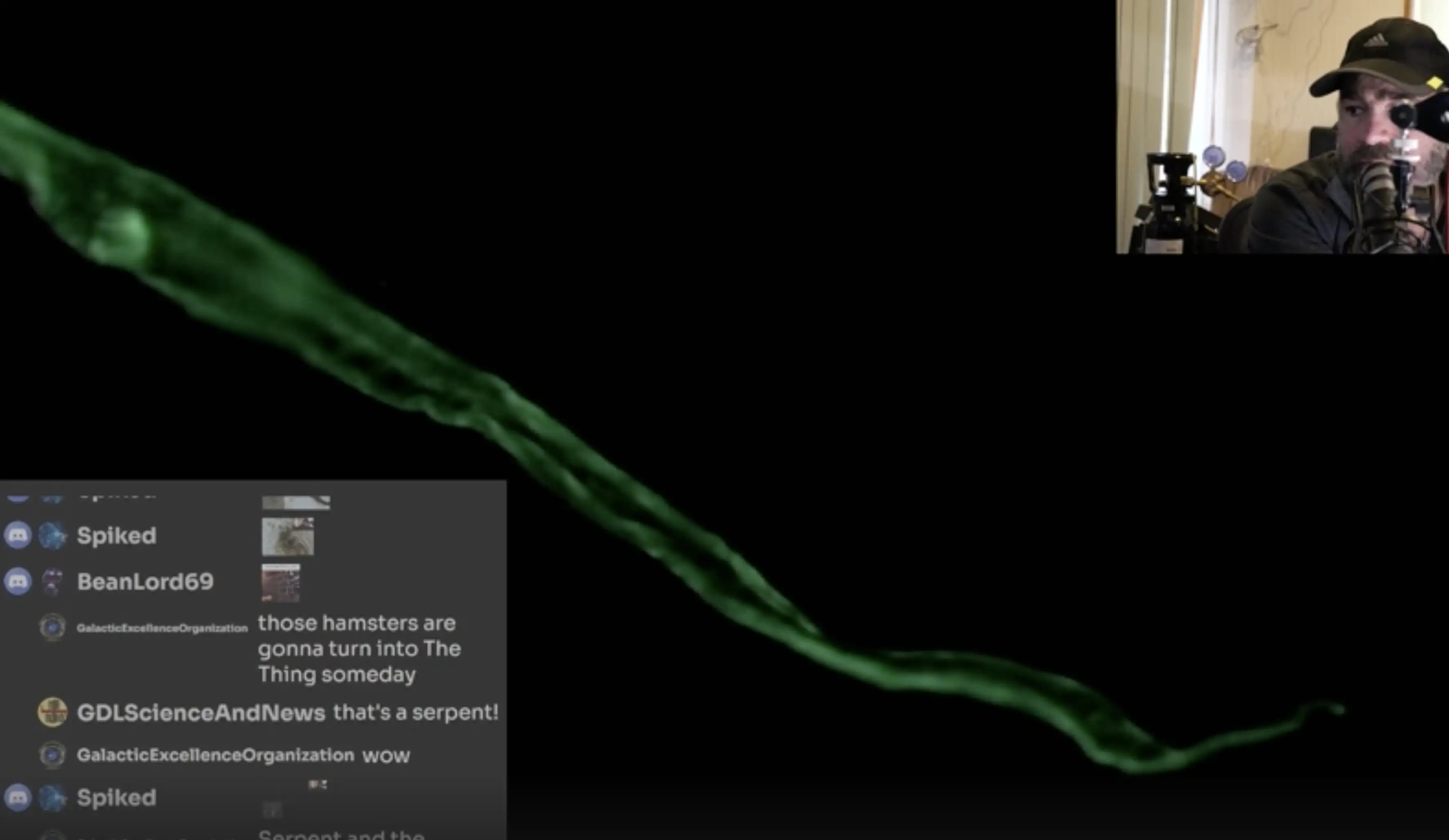
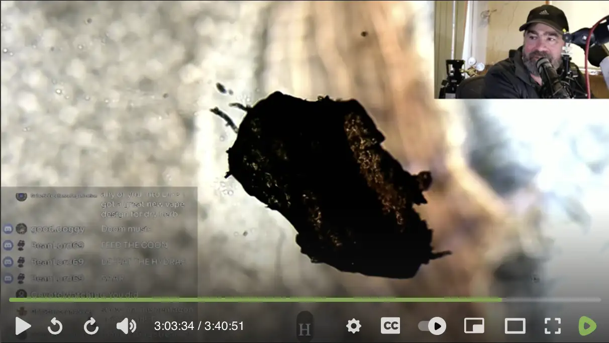
The next image shows the same fiber as the previous image, but under
a UV light, where the UV light excites the object to emit visible light
through fluorescence. In fluorescence microscopy the camera captures
only the intensity of the emitted light but not the hue, but the green
tint used in the image is arbitrary, and the image might as well be
displayed in grayscale or with some other tint. On his website McCairn
described the fiber as a "fibril", even
though in one of his presentation where he showed a version of the image
with a scale bar, the fiber was shown to have a diameter of about 15 µm:
[https://
The fiber in the image above has a fold that runs along the middle lengthwise. When I asked McCairn on his Discord what the fold was, he said: "And the fold running length wise is indicative of it being a coherent structure, that is one of the reasons why fibril is a fair description of the phenomenon. Again this is you running into the axiom of those that can and those that can't. This is a highly unusual amyloidogenic form." And when I said the structure was too big to be called a fibril, he said: "And one could say fibrillar forms, but fibrils is good enough at this stage, again because of the extremely large size differential between normal amyloids and these unusual fibrin amyloids." Then I asked someone else: "How do you even know that the structures shown by McCairn and Micronicle are fibrin clots? I think fibrin clots wouldn't have the kind of lengthwise fold in the middle as McCairn's green mystery fibril." But McCairn replied: "This objection to raw data comes from your years of extensive lab experience working with amyloids, I presume?" He also seemed to suggest that the mystery fiber he showed on his website was the same type of structure as the fiber in Mr. Micronicle's image (even though Mr. Micronicle's fiber didn't have a lengthwise fold running along the middle, and it didn't look as flat as the fiber on McCairn's website).
But anyway, are there even mini calamari clots that float freely in human blood, and that have a diameter of about 10-30 micrometers and a length of hundreds of micrometers?
The purpose of fibers of fibrin is to form a web that captures red blood cells, so then the tangled mess of fibrin and blood cells form a clot that blocks a punctured blood vessel. The reason why large clots have elongated shape is because they conform to the shape of a blood vessel, like if some entangled sticky mess was poured through a tube and the tube got clogged.
But if there's a micrometer-scale clot of fibrin that floats freely in the blood, why would it have an elongated shape? Did the clot form inside a small blood vessel and then get dislodged?
ChatGPT said:
1. Fibrin Clot Structure In Situ
Fibrin forms a mesh or network of thin fibers during clot formation, creating a gel-like matrix that stabilizes platelets and red blood cells.
This structure is highly entangled and irregular, not a single, uniform fiber.
2. Shape of Freely Moving Clots (Emboli)
When parts of a fibrin-rich thrombus dislodge and enter circulation, they become emboli.
The shape of these emboli depends on:
Origin: Venous thrombi are often elongated or serpentine, as they form in the low-shear environment of veins.
Mechanical deformation: They may elongate as they pass through blood vessels.
However, emboli are generally amorphous, irregular, or elongated masses, not slender, fiber-like threads.
3. Fiber-like Structures?
While fibrin fibers themselves are nanoscale or microscale strands, a free-floating clot is a bulk mass of these fibers entangled with cells and plasma proteins.
You will not see individual fibers moving freely in the bloodstream in vivo.
I asked McCairn that if the fiber in his microscope image formed inside a small blood vessel and then got dislodged, then why is it flat, and why does it have the lengthwise fold in the middle. He came up with this weak answer: "Good question why does this diseased protein have the form it does, it should be noted it's not the only form there are irregular & spheroid forms. I think it comes down to location of formation, nature of the underlying nano-scale fibril geometry, the species of protein, fibrinogen which makes long linear forms. Again we are in a process of discovery and relying on orthodox frameworks you constrain yourself to being able to competently describe it. But it's a common form, even described in the published literature." But I didn't find any paper in the medical literature that would've described fibrin clots as having a flat ribbon-like shape with a lengthwise fold running along the middle.
When I asked why McCairn's fiber has the lengthwise fold, ChatGPT said the fiber might be a fiber of cellulose where the hollow lumen in the middle has collapsed:
A lengthwise fold or groove is not characteristic of fibrin or amyloid aggregates. Instead, it is a well-known feature of certain textile or environmental fibers:
Cotton fibers: Typically ribbon-like, twisted, and often show central folds or convolutions due to the collapse of the hollow lumen inside the natural fiber.
Cellulose fibers (like paper fibers or lint): Also tend to flatten and develop longitudinal folds.
Synthetic fibers: Usually round or oval in cross-section, smooth, and don't typically have the central fold unless manufactured with a special cross-section.
Based on the ribbon shape, width (tens of micrometers), and the longitudinal fold, this looks much more consistent with a collapsed plant-derived cellulose fiber (e.g., cotton lint) rather than any protein fibril or clot material.
Added later: I now found the stream where McCairn originally
presented the microscope images of the fiber. [https://
At time 2:23:18 McCairn showed the fiber in UV mode and said: "That whole structure is fluorescing green. That tells me that it's highly likely that we're looking at an amyloid structure in the patient's sample." However he only showed the fiber after he had applied the ThT but not before it, so you couldn't see if the fiber was already autofluorescent without the ThT.
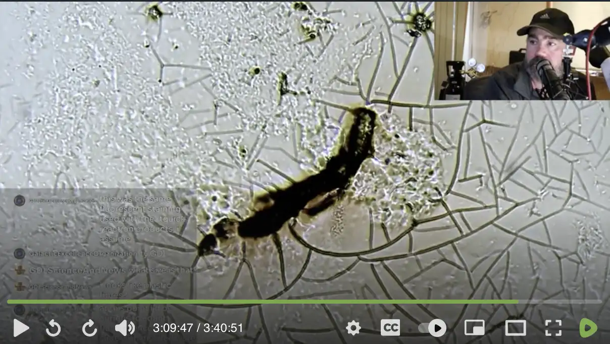
From the stream you can see that the end of the fiber has a torn appearance, which doesn't seem like characteristic of a fibrin clot:

You could also see that the diameter of the fiber tapers out towards the other end:
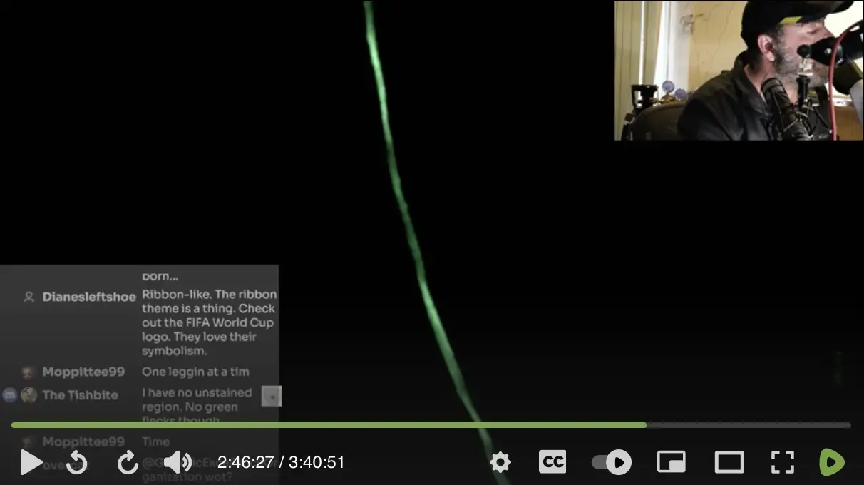
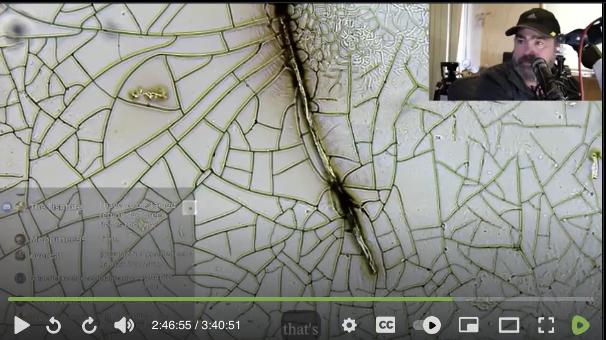
Next McCairn compared Lyndsey's blood to a sample of his own blood, where some regions of the slide was stained with ThT but others were not, so while he was panning around the slide, it was not always clear if he was looking at the stained region or not.
His slide contained this fiber that was fluorescent, even though I don't know if it was because of the ThT staining or not: [2:46:27]
His slide contained this structure that was fluorescent but that was not in plane with the rest of the sample, so McCairn said it's probably dust: [3:03:02]
His slide contained this fluorescent fiber, which was not in plane with the rest of the sample, so after he had focused the microscope on the fiber, he had to refocus the microscope to get the main part of the plane in focus, and McCairn said the fiber may have been dust: [3:07:14]
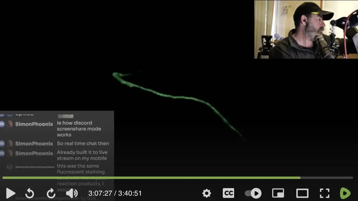
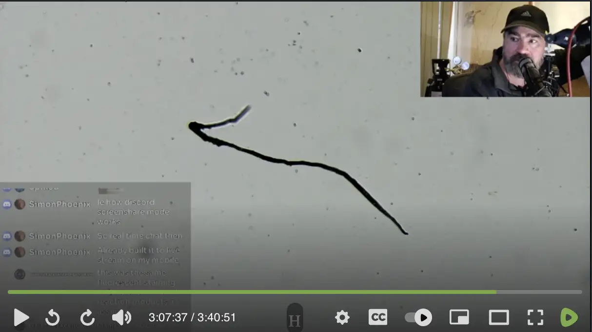
Next he showed this fluorescent fiber, which unlike the previous fiber was approximately in plane with the rest of the sample: [3:09:38]
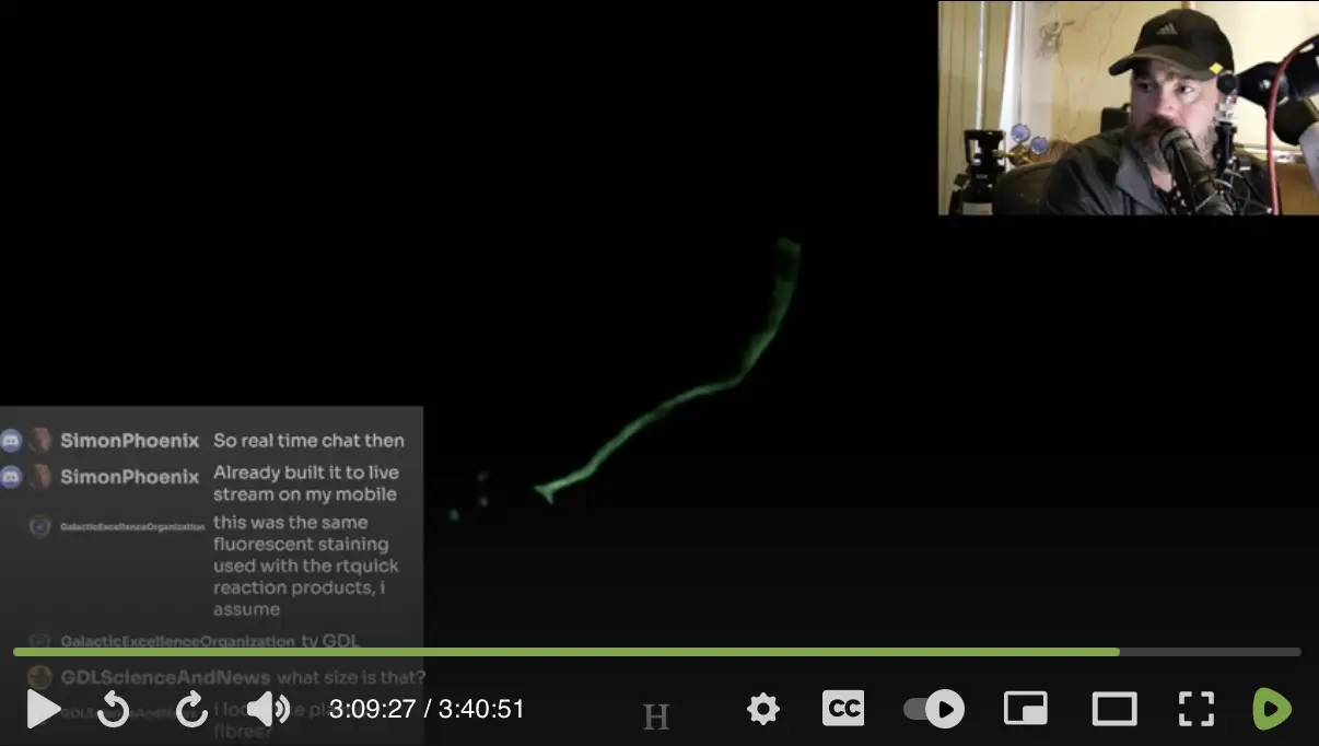
At time 3:16:19 when McCairn instructed his followers on how to send a blood sample to him, he said "don't put a cover slip on it, air dry it overnight" (but he didn't mention that would make it likely that dust would land on the slide while the blood was drying).
At time 3:17:23 he said: "I want to see how often I can find these structures, that are in plane with the blood, have a filamentous and helical stucture to them, and fluoresce." Some of the fibers that McCairn said were likely dust fulfilled 2 of his 4 criteria, because they fluoresced and had a filamentous structure. By "helical structure", he likely meant the kind of lengthwise twists that are called convolutions in cotton, so cotton fibers might easily fulfill at least 3 of his 4 criteria, even though if a cotton fiber from dust fell on the sample of blood after the blood already dried, I don't know if the fiber would be in plane with the blood so that the fiber would fulfill the 4th criterion.
McCairn published his second Substack post in May 2025. [https://
The post may have been partially generated by AI, because a heading before a list was formatted as Markdown. Substack doesn't even support Markdown, but some AI utilities add Markdown formatting to copied text:

McCairn's post has the air of cheap propaganda because of the
emphasis on harm done to babies. A common trope among morticians in alt
media is that they have a dramatic story to tell about dead babies, like
how the British funeral director Wesley said that there was about ten
times the normal number of babies dying, so the fridges were packed full
of dead babies. [https://
The focus of McCairn's post was on scary microscope images, which is reminiscent of content by the Quinta Columna and the Stew Peters crowd. ChatGPT said that McCairn's post "strongly resembles pseudoscientific or hoax material, exploiting scientific-sounding language and visual data without meeting the necessary standards of scientific proof".
In his post McCairn presented the images below, which both show the same fiber that he supposedly found in the 3-year-old's blood. The bottom image was taken under a UV light, where the UV light stimulates the Thioflavin T to emit visible light in the blue-green range. The microscope captures only the intensity of the light but not the color, so the green tint employed in the image is arbitrary. The bottom image shows that the fiber was fluorescent under a UV light even before McCairn had applied Thioflavin T on the sample, so McCairn said that the fiber was autofluorescent:

He also showed images of a second similar fiber, and he described the fibers as "autofluorescent fibrillar structures" and "UV-reactive fibrillar microclots" and "amyloidogenic fibrin microclots".
However just because McCairn's fibers are fluorescent under UV light doesn't mean that they are "amyloidogenic fibrin clots", but they might for example be fibers of cotton, because in the same way that a white t-shirt is fluorescent under UV light, fibers of cotton that have been bleached white are also fluorescent under UV light. (Based on the scale bar in the two images above, the fiber appears to have a diameter of about 5 µm, but later it turned out that the scale bar was incorrect and the diameter was actually about 15 µm, which fits within the typical range of diameter for fibers of cotton, which is around 12-25 µm.)
One of Greg Harrison's AI-generated documents had similar Markdown
bold formatting as McCairn's post. [https://
McCairn told me that his post was an AI-generated summary, and that he left "the markdown in for transparency". But I don't buy his excuse, because the Markdown formatting was not even a clear sign that his post was generated by AI, and he could've been much more transparent by explicitly writing that his post was an AI-generated summary.
The Markdown formatting appeared in the heading of a list titled
"Clinical Case Summary", so at first I
thought McCairn meant that only the list was generated by AI based on
the rest of the post, and therefore I was wondering why the list
mentioned details that were missing from the rest of the post. But
McCairn clarified that he meant that the entire post was an AI summary
based on a longer original article: [https://
McCairn supposedly does his blood analysis using slides of blood that
he receives in the mail from his followers, even though it's not clear
if that's how he received the sample of blood of the 3-year-old. I
pointed out that if mini calamari clots are so common that he frequently
finds them in small random samples of blood, there should be vast fleets
of mini calamari clots swimming around in the bloodstream of people. But
he refused to even answer me what volume of blood he analyzed: [https://


The Twitter user Markus pointed out that McCairn said one of his
microscope images was taken at a 4-fold magnification, but the scale bar
seemed to indicate that the image was taken at a much higher
magnification level: [https://

ChatGPT said that typically "the field of view (FOV) for a 4x objective on a standard microscope with a 10x eyepiece is around 4-5 mm (4000-5000 µm)", but based on McCairn's scale bar, the area shown in his image was only about 550 µm wide, which is only about 12% of the typical FOV:

But McCairn said he didn't even use an eyepiece, so the FOV at 4x magnification should be about 40-50 mm, which means that the visible width of his image is only about 1.2% of a typical FOV.
When I asked ChatGPT if an eyepiece can go up to 100x, it said 100x eyepieces are extremely rare and not practical for most applications, and typically eyepieces only go up to about 15x or 20x magnification. When I asked what could explain McCairn's narrow FOV if the image was taken without an eyepiece, ChatGPT wasn't able to give any reasonable explanation. But it did say it was possible that the scale bar was wrong or the magnification level was reported incorrectly.
McCairn replied to me: "The field of view is not
0.5mm you cretin, the scale bar shows the pixels derived from the camera
using a calibration slide placed on the slide holder. you are looking at
an amyloidogenic fibril that is 100's of micrometers in length and the
scale bar in red shows 50 micrometers not 0.5mm you moron". [https://
In the last tweet I quoted above, McCairn referred to an SEM image of
the 3-year-old's fiber that he had now posted on Substack. His SEM image
was taken at 40-fold magnification, but the object in the image now took
up about 50% of the FOV: [https://

So why did the same object take up about 75% of the FOV in the other image that was supposedly taken at 4-fold magnification?
The object in the new SEM image is about 731 pixels wide, and the 1 mm scale bar is about 460 pixels wide, so the width of the area covered by the object is about 1.59 mm. But in the earlier optical microscope image, the width of the area covered by the object was only about 0.38 mm based on the scale bar.
Jikkyleaks asked: "Kevin are your scales correct
on the SEM pic? In the first picture it looks like that fibril is about
500micrometres long, but the SEM scale is marked in millimetres."
[https://
But anyway, I still didn't have an answer to why the old microscope
image had a FOV of only about 0.55 mm, even though the image was
supposedly taken at 4-fold magnification. So I asked McCairn: "So was your 4x image cropped or not? If so then what
percentage of the width of the original image did the cropped image
show? It's probably not nearly enough to explain why the FOV was only
about 0.55 mm even though a typical FOV at 4x magnification would be
about 40-50 mm." [https://

McCairn seems to routinely screw up the scale bars in his microscope
images. In this image the scale bar is shown to be 10 µm wide, even
though it's about 13 red blood cells wide, and red blood cells have a
diameter of about 7 µm, which means that the scale bar is really
somewhere around 90 µm wide: [https://

McCairn responded "Yes the scale bar is wrong it
should say 50 micrometers, the 10 micrometers is a holdover from a scene
done for more detailed analysis for an individual who wanted their blood
looked at." [https://
I have joined Kevin McCairn's Discord server multiple times since 2020, and I was even made into a moderator on the server at one point, but I have usually left the server after a while because it had a very low quality of posts. McCairn banned me from the server in May 2025, because I said that the calamari clots were fake and he was probably controlled opposition.
Since his early videos in 2020, McCairn has said that he got COVID from the Korean superspreader event in Daegu, and that he had severe neurological symptoms, which was a reason why he knew that COVID had a neurotropic effect and he decided to focus on researching the neurological aspects of COVID.
However the problem with his story is that the Korean superspreader event occurred in February 2020, but McCairn says that he was in Daegu from late November into December 2019. I hadn't seen anyone call him out for his claim until I brought it up on his Discord:

I used to think McCairn probably had some other illness but he mistook it for COVID, but after I found out how there's also many other people in alt media who claim that they were among the first people in their country who got COVID, I started to think it's possible that he just made up the whole story about having COVID.
It also seemed like an unlikely coincidence that McCairn was an early YouTube streamer who was focused on COVID, but he also happened to be at the right place at the right time to get infected with COVID very early on. Often if a fabulist invents an embellished biography for themselves, they insert themselves in various locations around the world at a time when some event of historical importance happens to occur at the location. But the Korean superspreader event was probably the best-known event that occurred in Korea in the entire year of 2020.
In the unlikely scenario where McCairn actually got COVID in November or December 2019, he might have been the first person with COVID in Japan, because he claims that he was still sick with COVID after he returned home to Japan in December 2019.
In the screenshot above, I pointed out how on January 28th 2020 UTC,
Steve Pieczenik claimed that a month ago he had the first case of COVID
in the United States, and he got COVID after he met with a Chinese
student from Wuhan (because I guess he didn't count
the student as an earlier case of COVID). [https://
I don't know if McCairn actually even visited Daegu in 2019 like he
claims, but in one video he also said: "I got hit by
the biggest superspreader event in Asia, right early on, in November to
December." [https://
McCairn's American sidekick Charles Rixey also claims that he got
COVID at a time when there were only a few reported COVID cases in the
US: [https://

Added later: I didn't find the early videos on McCairn's YouTube channel archived anywhere, because his first channel was deleted fairly early on, and his videos were not even archived by AltCensored. But these were McCairn's earliest videos about COVID I found that were still available online:
I listened to a couple of the videos above, and I searched through
the transcripts of a few more videos, but McCairn only said he may have
gotten COVID in Daegu in one of the videos, which was a video he did
with Addy Adds in March 2020. In the video McCairn said: "And, uh, where I had my lab, which was in South Korea in
a city called Daegu, they'd asked me to sort of come and give a, uh, to
help them in a project they were doing. And that time was October, no,
sorry, November - I want to say it was November into December, but late
November, right. And at that time, um, I came back, and over the
Christmas period, I got really, really ill, right. And, um, it's sort of
left - like the fever I was delirious, so I can't - and again, so I get
memory issues because of the head injury. And so apparently when I was
in the fever bit, I was coming downstairs and thinking days had passed.
And, uh, it, it wasn't, you know, and it took me a week to sort of get
over that. And then I was left just, um, with, it's called dyspnea where
you can't really breathe." [https://
I have pointed out to McCairn that individual fibrils of fibrin have a string-like shape because their purpose is to form a web that traps red blood cells. And large blood clots have an elongated shape because they conform to the shape of blood vessels. But that doesn't mean that if there is an intermediate-scale formation of fibrin that floats freely in the blood, like the microclots McCairn supposedly keeps finding in his blood samples, the formation would also have an elongated string-like shape, unless for example the formation first develops inside a small blood vessel and then get dislodged from the vessel.
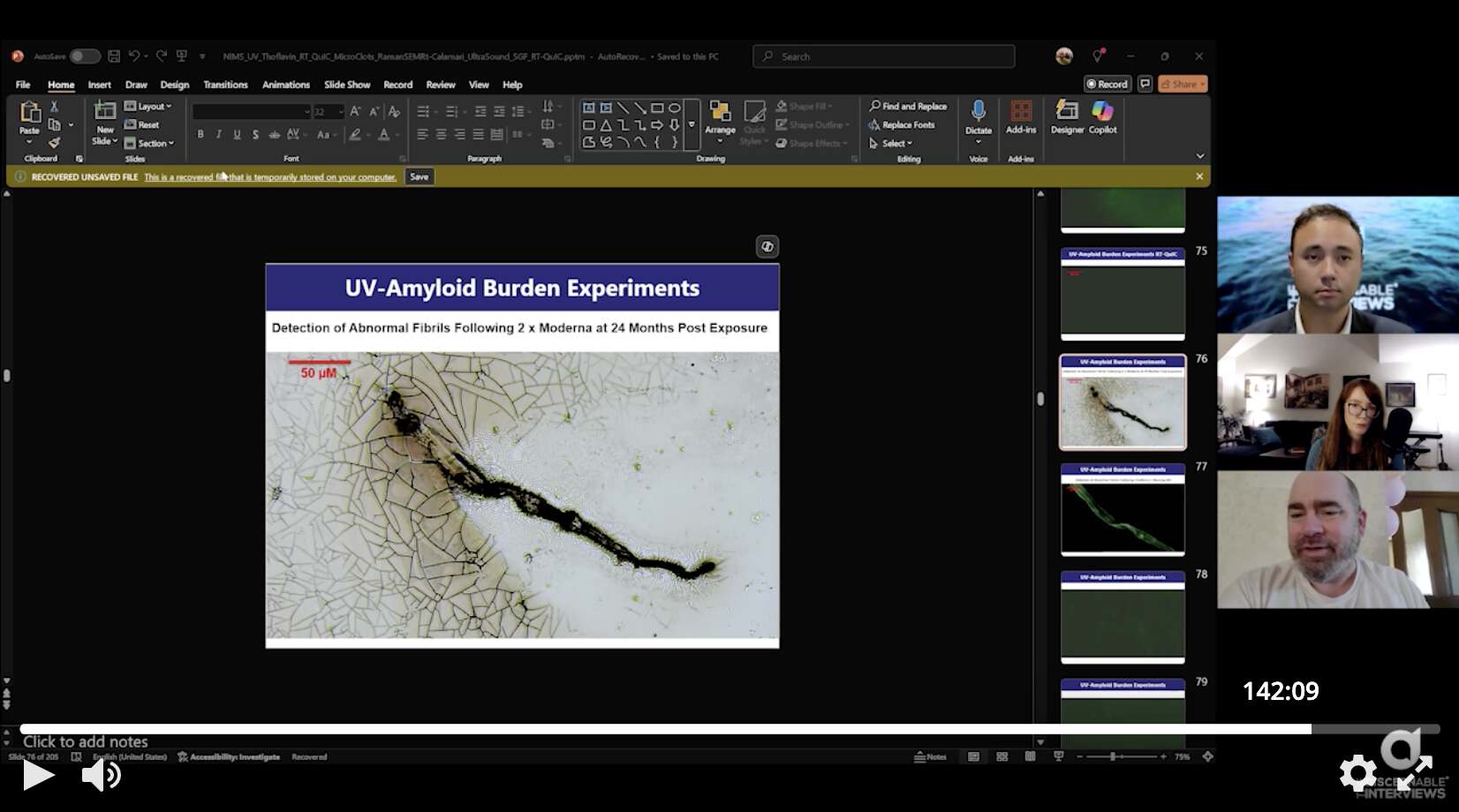
In McCairn's first Substack post about the 3-year-old's fiber, he
didn't attempt to explain why the fiber would have an elongated shape if
it was a fibrin clot like he claimed. But now in his second post about
the fiber, McCairn provided the following explanation for why the fiber
had a string-like shape: [https://
These comparisons between nano- and macro-structures highlight the conserved geometry of pathological amyloidogenesis. This scale-invariant preservation of fibrillar architecture aligns with prior biophysical studies demonstrating that amyloid formation follows universal thermodynamic pathways, forming twisted ribbon-like or lamellar structures irrespective of protein species or environmental origin (Chiti & Dobson, 2017; Eisenberg & Sawaya, 2017). The similarity across scales - from nanometer-thick fibrils to centimeter-scale clots - suggests a deeply encoded biophysical template likely seeded by persistent amyloidogenic peptides, such as SARS-CoV-2 spike protein.
However I'm not convinced by McCairn's explanation, and I didn't find any part of the two papers he cited that actually backed up what he wrote, or that would've explained why amyloid formations on the micrometer scale would have a string-like shape.
When I asked ChatGPT to describe the shape of micrometer-scale amyloid formations in the human body, it gave me the following list:
1. Aβ (Alzheimer's)
- Shape: Dense plaques (spherical, irregular, starburst)
- Size: 20-200 µm
- Location: Extracellular (brain cortex, hippocampus)
2. Tau (Alzheimer's, FTD)
- Shape: Neurofibrillary tangles (flame-shaped, globose)
- Size: 10-50 µm
- Location: Intracellular (neurons, dendrites)
3. α-Synuclein (Parkinson's, LBD)
- Shape: Lewy bodies (round with halo), neuritic threads
- Size: 10-25 µm
- Location: Intracellular (neuronal soma, axons)
4. Transthyretin (TTR, Amyloidosis)
- Shape: Linear, patchy, or amorphous deposits
- Size: 10-100 µm
- Location: Extracellular (heart, nerves, GI tract)
5. Light Chains (AL Amyloidosis)
- Shape: Nodular, perivascular linear deposits
- Size: 20-200 µm
- Location: Extracellular (kidneys, heart, tongue)
6. IAPP / Amylin (Type 2 Diabetes)
- Shape: Sheet-like or intercellular ribbon deposits
- Size: 10-50 µm
- Location: Extracellular (pancreatic islets)
7. Serum AA (Systemic Amyloidosis)
- Shape: Diffuse, collagen-bound linear aggregates
- Size: 10-100 µm
- Location: Extracellular (liver, spleen, kidneys)
8. Fibrin-Amyloid (Microclots, Long COVID)
- Shape: String-like, thread or net-like filaments
- Size: 50-500 µm
- Location: Circulating blood, capillaries, microvessels
ChatGPT said that IAPP/amylin forms "sheet-like or intercellular ribbon deposits", but IAPP doesn't quality as a formation with a diameter on the micrometer-scale, because ribbons of IAPP typically have a diameter of about 10-15 nanometers. Out of the 8 types of formations listed by ChatGPT, only the microclots associated with COVID had both a string-like shape and a micrometer-scale diameter. But I couldn't get ChatGPT to cite any source which said that microclots clots associated with COVID actually had a string-like shape, so ChatGPT may have been influenced by McCairn's Substack post which I had shown to it earlier.
I asked McCairn: "Is there some specific part of
the papers you cited which says that formations amyloid protein have a
tendency to form into a ribbon-like shape in the micrometer
scale?" [https://
Here we go again, you are not looking at the canonical amyloidogenic form, merely based on the size of the aggregations .
How many times do I have to tell you this? That is why I as the domain level expert uses the language as I see fit.
Especially when it reacts to all the diagnostic measurements used to detect amyloids.
ThT Fluorescence, SEM structural characteristics, RT-QuIC reactivity, Raman Spectra confirmation.
I replied: "You suggested amyloid structures have a 'scale-invariant preservation of fibrillar architecture' so they have a tendency to form into a ribbon-like shape on the micro-scale, and you cited two papers from 2017. Which part of the papers supports your point?" But he didn't answer me.
A paper by Pretorius and Kell featured the images of microclots
below, which had a globular or blob-like shape, but they didn't look
anything like the fibers that McCairn finds under a microscope: [https://

I asked Douglas Kell if his group had seen the kind of fiber-like
microclots that were shown in McCairn's Substack post, but he just
replied "these experiments are quite different so
comparisons are not usefully made". [https://
Then McCairn wrote: [https://
In the experiments conducted by @dbkell and colleagues, the whole blood is spun in a centrifuge, larger aggregates would be pulled to the bottom of the tube.
Spinning and looking at the plasma phase would leave the smaller amyloidogenic seeds in the plasma phase. This allows automated sorting through techniques like flow cytometry. This is a better approach for batch processing, you will miss the larger aggregations due to centripetal forces being greater on larger amyloidogenic forms.
Slide analysis is much slower, requires someone with trained eyes, is labor intensive, and requires the follow on tests of ThT staining, SEM/EDX, Raman Spec, for categorization.
However if vaccinated people are now suffering from a novel pathology where their blood is full of clots that look like textile fibers, then is there anyone except McCairn who has published an image of one of the clots? I didn't find any similar images of clots published by Pretorius and Kell. Similar images of fibers in blood have been presented by Ana Mihalcea, Shimon Yanowitz, David Nixon, people at Burkhardt's pathology conference, and Mr. Micronicle, but as far as I know, none of those people claimed that the fibers were clots made of fibrin.
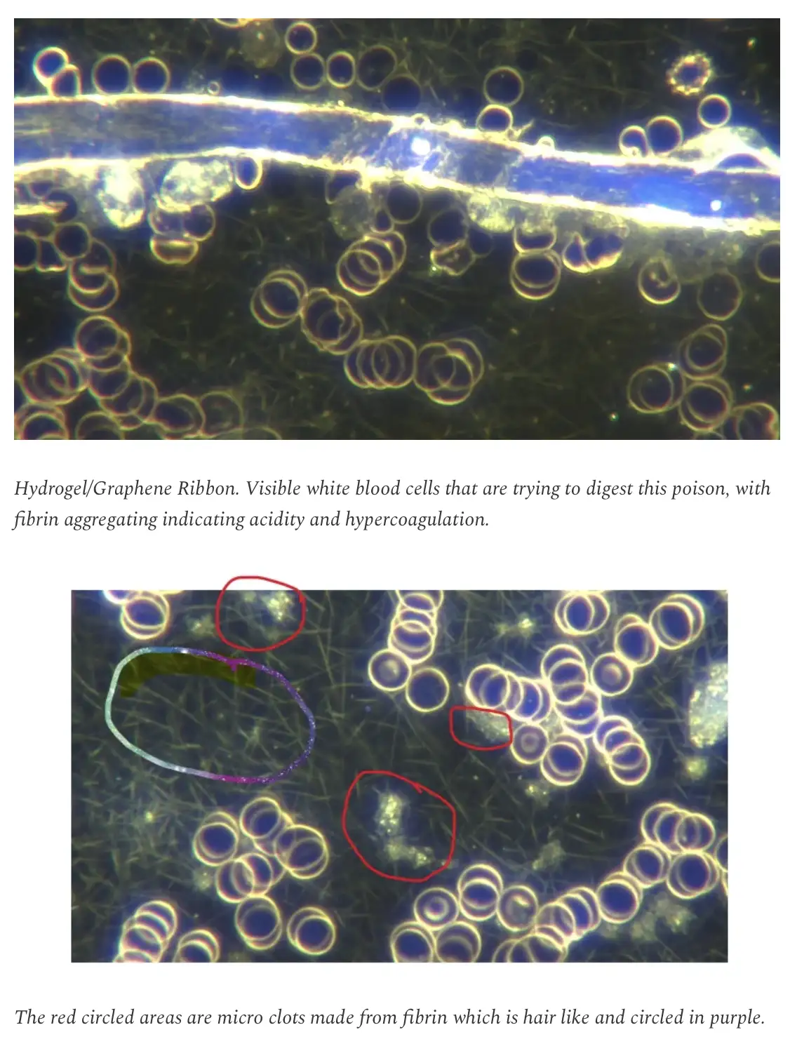
Mihalcea said that in her microscope images of blood, a web-like
structure was fibrin, and blobs within the web were fibrin microclots,
but a larger isolated fiber was a "hydrogel/graphene
ribbon": [https://
(Added in 2026: I now found one Substack post
where Mihalcea said that a fiber with a diameter of about 30 µm was a
fibrous rubbery clot "in early stages". [https://
Added in June 2025: I now asked McCairn again that when he cited the
two papers from 2017, what part of the papers supported his claim that
aggregates of amyloid protein tend to organize into a ribbon-like shape
on the micrometer scale. [https://
These β-structured oligomers are able to grow further by self-association or through the addition of monomers, often with further and sometimes dramatic structural reorganizations, to form well-defined fibrils with cross-β structure and a high level of structural order (Figure 1). Alternatively, the disordered aggregates or native-like aggregates can grow without any major structural conversion and give rise to large amorphous deposits or native-like assemblies, respectively, retaining the structure characterizing the initial oligomers (Figure 1).
Such large aggregates, including amyloid, amorphous, or native-like assemblies, have links with human disease as they accumulate in well-defined pathological states. Tables 1 and 2 list the proteins and the disorders that have now been identified to be associated with the formation of amyloid fibrils or other types of aggregates, respectively. (Supplemental Tables 1 and 2 also list, for each protein, references reporting the identification of the protein in the aggregates and the characteristics of the aggregate type.) We have arranged both tables in terms of proteins rather than disorders to stress the fact that many of these proteins have been found to be involved in a variety of pathological conditions. Interestingly, immunoglobulins or their subunits are found in all the different types of protein aggregates, including amyloid (as in light-chain amyloidosis), amorphous (as in light-chain deposition disease), and native-like (as in Berger disease) structures, thus representing a remarkable manifestation of the multiplicity of pathways existing in protein aggregation and of the structures and morphologies that can be generated (24-26). Those proteins that form intracellular inclusions of types that are still debated, such as TDP-43 and p53, are included in Table 2 with a footnote explaining this uncertainty.
But I pointed out that Figure 1 illustrated typical nano-scale amyloid fibrils, and not micrometer-scale amyloid formations. And in Supplemental Table 1, the shape of various types of amyloid structures was characterized as "intrinsically disordered":

Then McCairn replied:
You're so retarded! This particular amyloid, is novel and not listed because you're looking at science being done in real time.
The principles of misfolding though, are likely the mechanism leading to the macroform and of course the species of protein undergoing change. Fibrin is inherently primed to make macromolecular structures, hence the amyloidogenic form is going to be larger.
Which is why I have gone to the effort of showing concordant structure across scales.
So I told him: "Post-COVID clots are not relevant to my question. You cited two pre-COVID papers to support a claim that amyloid aggregates have 'scale-invariant preservation of fibrillar architecture' and they form 'twisted ribbon-like or lamellar structures irrespective of protein species'." And McCairn replied: "That's because there is no post-COVID manuscripts showing the phenomenon, in the manner that I have, you're looking at it being made right now, and disseminated to the public. It is usual in scientific writing to point to key historical citations that demonstrate concordance within hypothetical frameworks." But he still didn't answer my question, so I told him: "We already know you claim that baby calamari clots have a tendency to form into a ribbon-like shape on the micrometer scale. The question was which part of the sources you cited supports the claim that the same applies to other types of amyloid formations."
But then McCairn said "there is a coherency in
the epistemological grounding of the amyloid PRION formation", so
I told him that it sounded like great pseudo-profound bullshit, because
the pseudo-profound bullshitters are always talking about epistemology:
[https://


DopplerEffect93 is a user on Twitter who has a PhD degree in
neuroscience, and who worked on a postdoctoral project that involved
analysis of amyloid fibrils: [https://
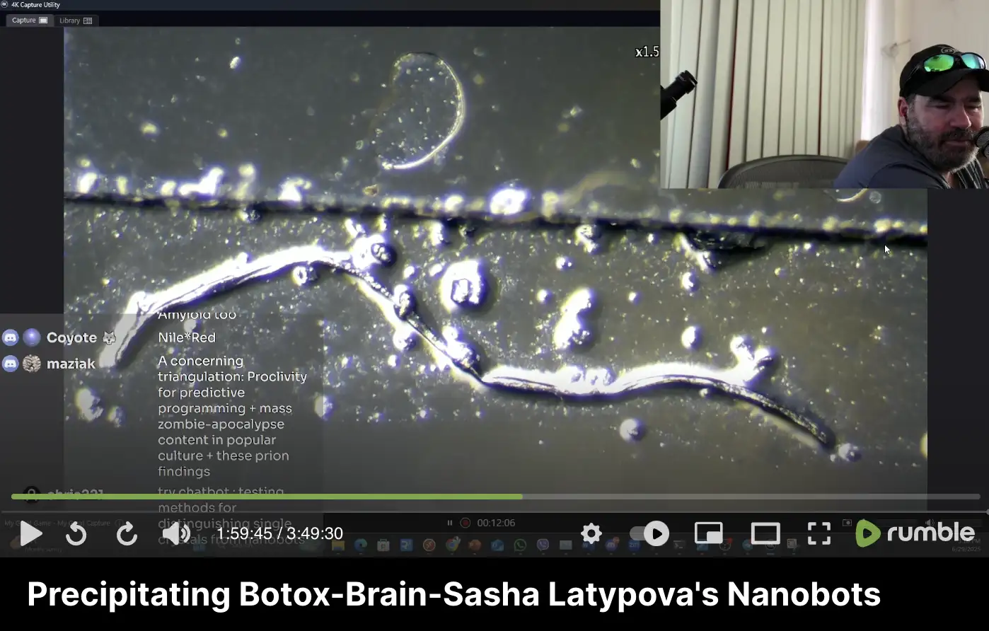
He said that the string-like object in McCairn's microscope image was
too big to be an aggregate of amyloid protein, and the object looked
like dust or debris. McCairn cited a paper by Pretorius and Kell which
said that "the fibrinaloid microclots that we
observe are typically in the range 1-200 µm on their longest
axis": [https://

However the text he quoted referred to this figure, which showed that the microclots described by Pretorius and Kell did not have a string-like shape:

Added in June 2025: CHD posted a video where Suzanne Humphries said
that when she looked at the contents of a vaccine vial under a
microscope, she saw structures that looked like circles and squares, and
they transformed into structures that looked like circuit boards: [https://

Under replies to the tweet, some random users posted about microscope images that they apparently thought were related to the discovery by Humphries. They included images of a chip-like structure shown by people in Burkhardt's pathology conference, graphene disks found by Zandre Botha, and the amyloid fibril that McCairn supposedly found in the blood of the 3-year-old (but a common denominator between Burkhardt, Botha, and McCairn is that they have all presented a laboratory analysis of the calamari clots):


The tweet about McCairn's amyloid fibril was posted by a user called
PinkBeachGirl1, which looks like a bot. It has posted almost 200,000
tweets: [https://

Added in June 2025: McCairn now did a stream where he looked at the
contents of a Moderna vaccine vial under a microscope. He saw these
fiber-like structures that were autofluorescent under UV light, and he
said "If I saw that in blood, I'd be like, uh, that
looks suspect": [https://
He also found this strongly autofluorescent fiber in the sample:
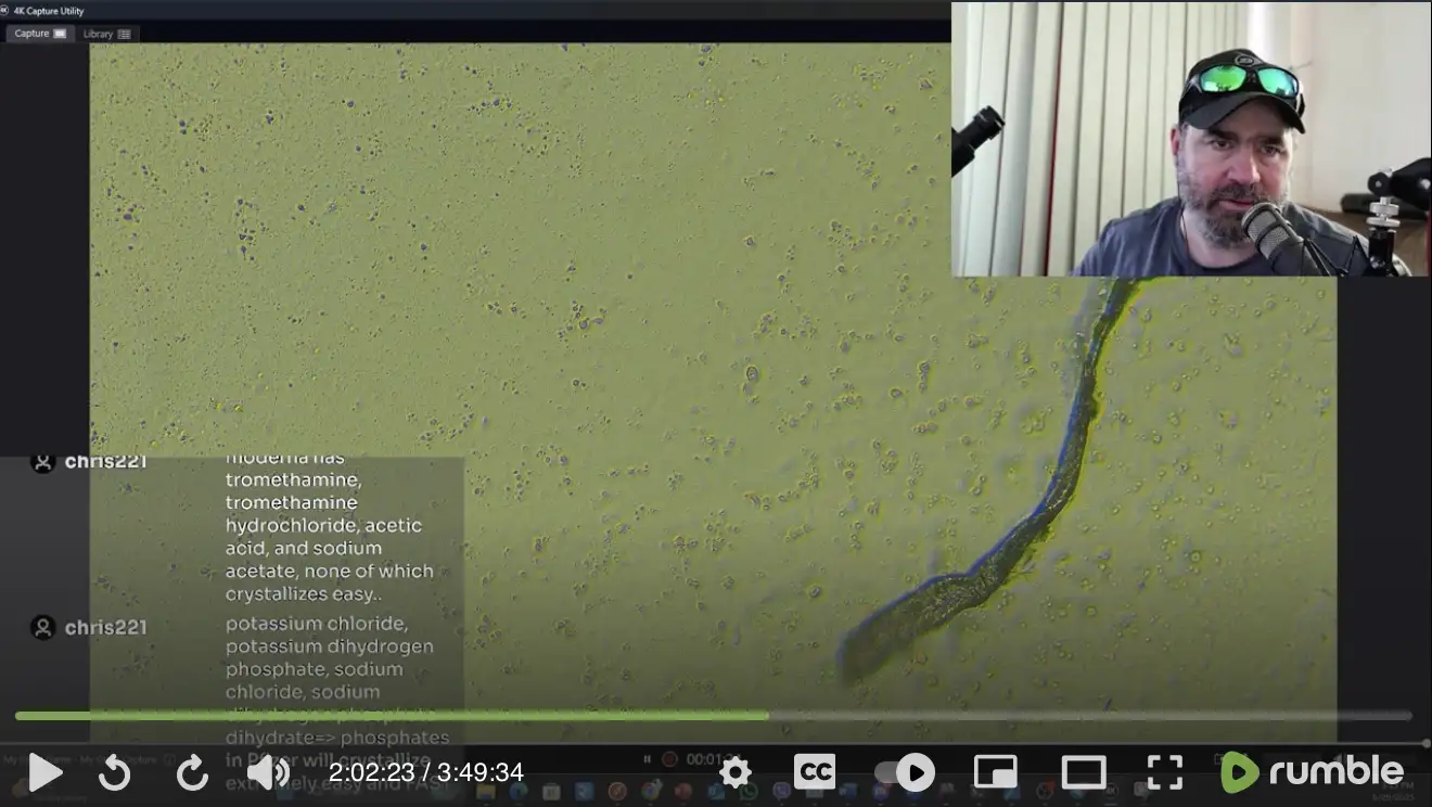
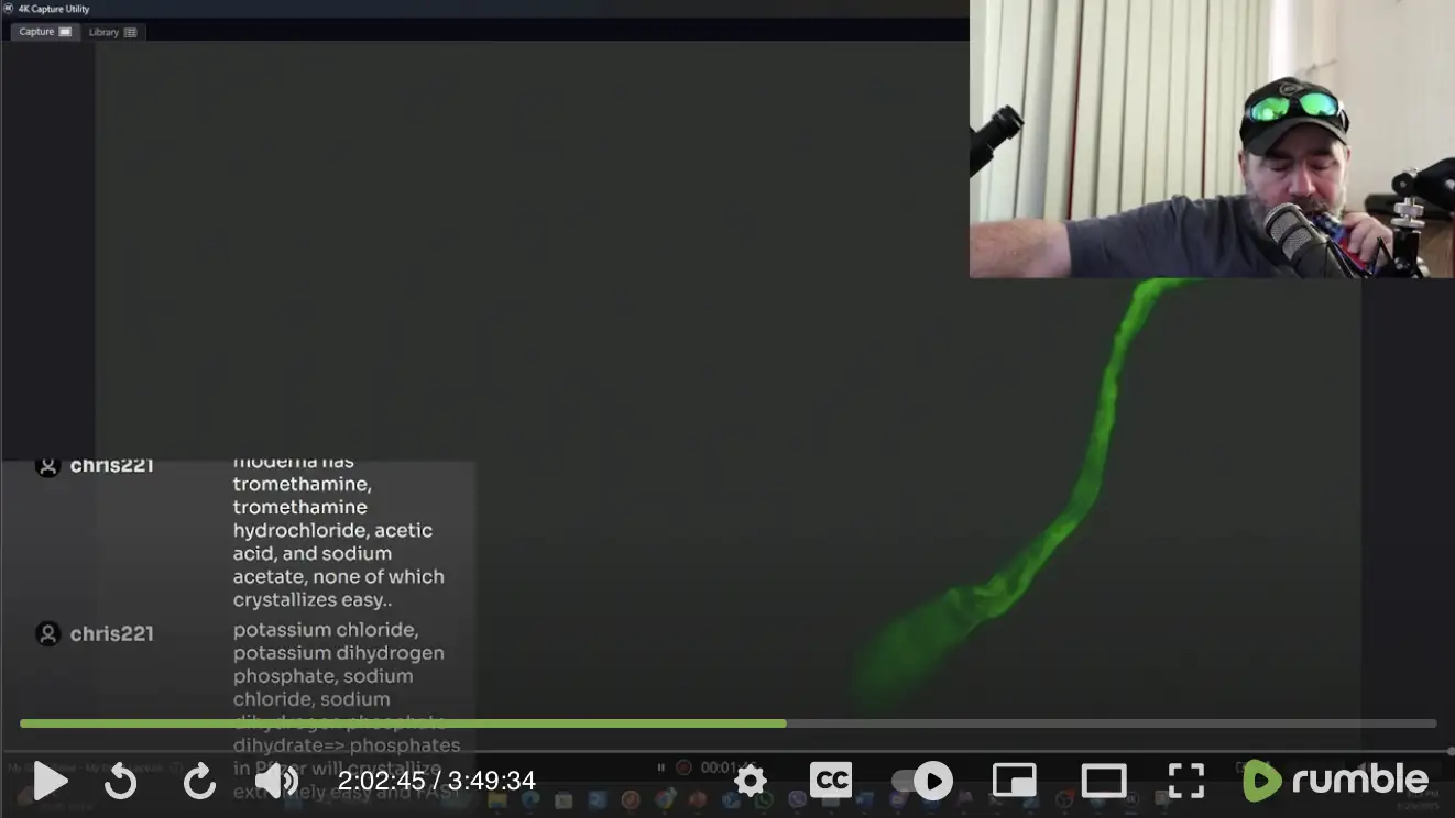
And this is another strongly autofluorescent fiber he saw:
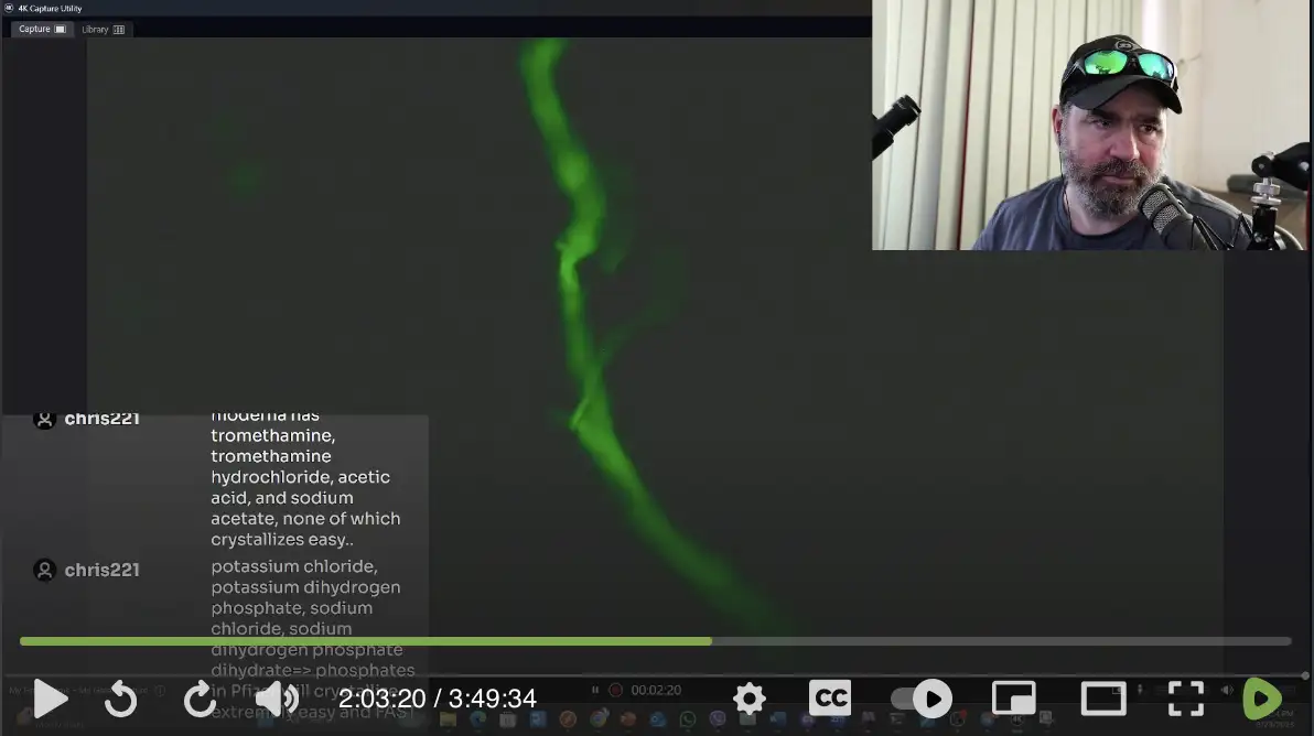
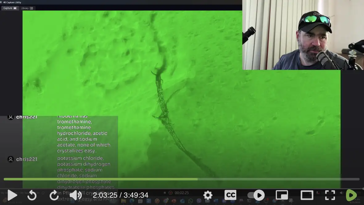
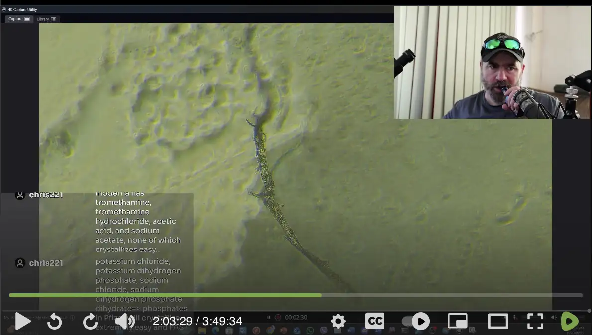
He said he didn't know what the fluorescent fibers were, but he said: "And the problem with there being bacterial contamination - I don't know how much those fibrous mats are like bacterial hydrogels in this instance." [2:05:23] He sounded like Ana Mihalcea when he suggested that random fibers he saw under the microscope were "hydrogel".
When I asked what bacterial hydrogels were, an LLM said: "Bacterial hydrogels (also known as Engineered Living Hydrogels) are a new class of 'smart' materials made by encapsulating living bacteria within a 3D water-swollen polymer network." Grok said:
Bacteria do not produce autofluorescent fibers with diameters of 10-50 micrometers that can be characterized as hydrogels. Typical bacterial extracellular structures, such as curli amyloid fibers or bacterial cellulose nanofibrils, are much smaller, usually in the range of 5-100 nanometers in diameter. These nanoscale fibers can form networks within biofilm matrices that behave like hydrogels due to high water content, but the individual fibers themselves remain far too thin to match the described size.
Bacterial cellulose, produced by species like Komagataeibacter, consists of nanofibrils that occasionally assemble into larger mats or pellicles, yet even bundled structures rarely approach 10 micrometers in diameter. Autofluorescence can occur weakly in some bacterial proteins or amyloids, but it is not a prominent feature of large fibrous structures. Larger hydrogel-like materials involving bacteria are usually engineered systems, where bacteria are encapsulated in synthetic hydrogels rather than naturally secreting thick autofluorescent fibers.
Below the first image shows a fiber without ThT staining and in non-UV mode, and the second image shows the fiber with ThT staining and in UV mode. McCairn showed that the fiber glowed brighter than the background after the staining:
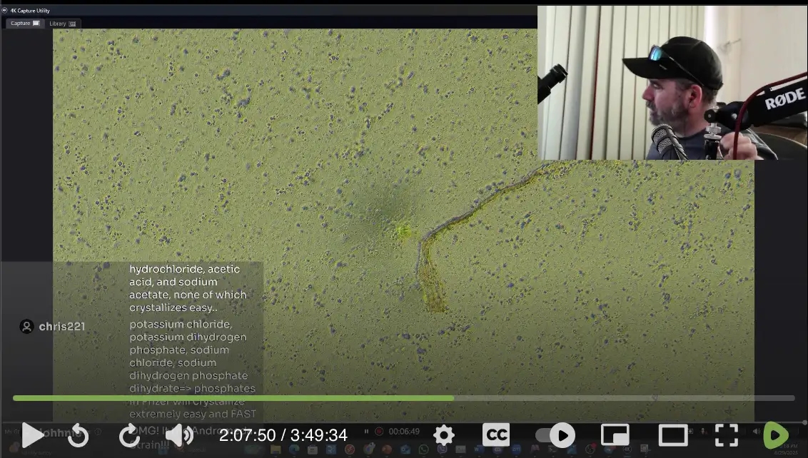
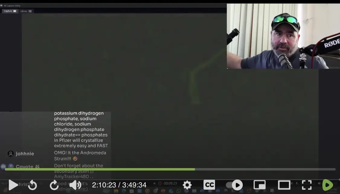
However he didn't menton that a few minutes earlier when he panned past the fiber, you could see that the same fiber was already autofluorescent before he did the ThT staining, so the fluorescence was not even because of the ThT:
At time 2:23:21 he said: "I can't explain the takeup of Thioflavins. So, well, the only thing I could think of was, well, these are bacterial." But he didn't mention that the same fiber that supposedly took up Thioflavin was already autofluorescent even without the ThT.
He also showed this fiber in the Moderna vaccine sample, which might be some textile fiber because it has a fairly uniform diameter of about 2-3 red blood cells (but the structures towards the bottom left side are not connected to the fiber):
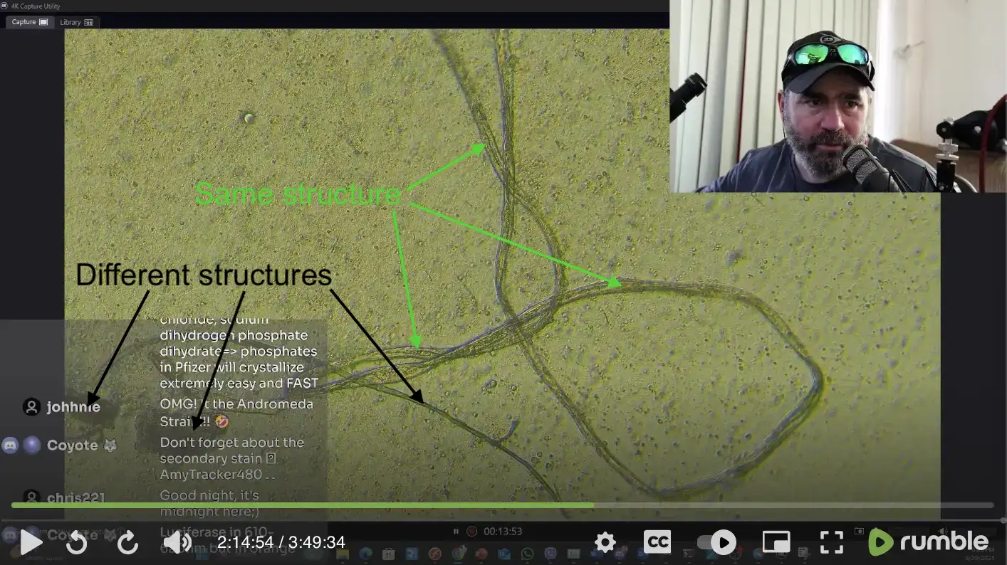
McCairn's slide included multiple different types of fibers and other structures, some of which may have been fungal or bacterial. Many bacteria and fungi glow in UV light, so UV lamps are specifically employed to detect bacteria or fungi on the surfaces of a room, or to see if a room has been cleaned properly.
The so-called "amyloidogenic fibril" that
McCairn found the blood of the 3-year-old may have also been a false
positive, because it was similarly autofluorescent even without ThT
staining: [https://
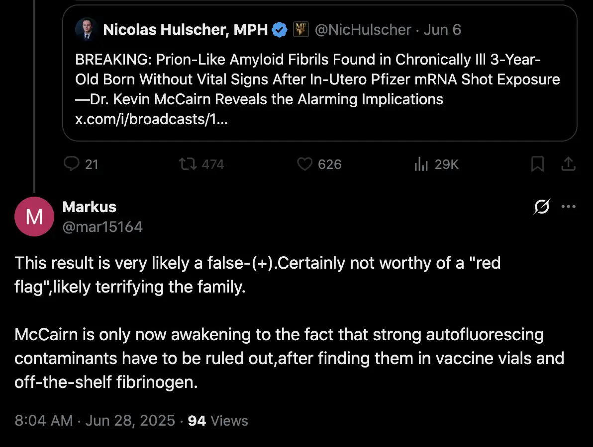
McCairn's new discovery reminded me of the mysterious fibers that
were supposedly discovered in a Moderna vaccine vial by Carrie Madej:
[https://

A biologist user on Twitter pointed out that amyloid fibrils are
nano-scale structures, and that McCairn's fiber looked like a fiber
typically seen on slides: [https://

In a video in 2022, McCairn showed the microscope image by David
Nixon below, and McCairn said: "Everything that I
see here is cholesterol, and literal fibers that you pick up in dust in
the air. What I've seen these people do - they're not doing it in clean
rooms - and you've gotta be in exceptionally clean rooms to make sure
that slides are not getting contaminated. Sorry. You don't get to say
that these are graphene internet fiber optic cables." [https://

McCairn's Substack post was titled "Amyloidogenic Fibrils in a Post-Gestational Case of mRNA Vaccine Exposure: Structural, Pathophysiological, and Biosecurity Perspectives". So the title didn't employ the term "amyloid fibril", which is a standard scientific term that refers to nano-scale structures, but the title rather employed the nonstandard term "amyloidogenic fibril", which might plausibly refer to something other than the typical meaning of an "amyloid fibril". In the post McCairn also referred to the so-called "amyloidogenic fibril" as an "amyloidogenic fibril formation", a "fibrillar formation", a "fibrillar aggregate", and a "macro-fibrillar form".
However Nicolas Hulscher's tweet about the post said "BREAKING: Amyloid Fibrils Found in 3-Year-Old After
In-Utero mRNA Injection Exposure". Hulscher also employed the
term "amyloid fibrils" in the title of his
Substack post: "BREAKING: Prion-Like Amyloid Fibrils
Found in 3-Year-Old Born Lifeless After In-Utero Pfizer mRNA Injection
Exposure". [https://
The junk amplifier accounts SenseReceptor and toobaffled also said
that McCairn found "amyloid fibrils" in the
sample of blood: [https://


McCairn linked to a ChatGPT conversation where he uploaded his images
of the 3-year-old's fiber, and he asked if the fiber conformed to "known amyloidogenic fibrillar forms of fibrin".
[https://
The diameter of the 3-year-old's fiber is about 10-20 µm based on the scale bar in the SEM image. When I asked ChatGPT what the diameter of amyloid macrofibrils was, it said the diameter was typically 50-200 nm:
Amyloid macrofibrils are higher-order bundles of smaller amyloid fibrils. Their diameter depends on both the protein type and the aggregation environment, but in published measurements:
- Single amyloid fibrils: ~6-12 nm diameter (core β-sheet cross-section)
- Protofibrils: ~2-5 nm (precursors before mature fibril)
- Macrofibrils: typically 50-200 nm, but sometimes up to 500 nm in thick bundles seen in plaques or clot-associated aggregates
For example:
- Alzheimer's Aβ plaques: macrofibrils often 80-150 nm
- Serum amyloid A (AA amyloidosis): bundles 50-200 nm
- Amyloid-rich fibrin(ogen) clots: ribbon-like macrofibrils up to a few hundred nm wide, occasionally merging into micron-scale sheets
In his Substack post, McCairn also wrote that the 3-year-old's fiber
was a "macro-fibrillar form". [https://
In McCairn's second Substack post, he showed optical microscope
images of the 3-year-old's sample of blood, and he indicated that he was
next going to run SEM, EDX, and Raman on the sample: "Guardians were explicitly briefed on the scope and
purpose of data collection, the nature of imaging techniques employed
(including UV fluorescence microscopy, Thioflavin T
staining, preservation for SEM/EDX analysis, and Raman
Spectroscopy), and how the data would be analyzed and
disclosed." [https://
However in his third Substack post, McCairn only posted SEM images of
the sample, but he didn't describe the results of EDX or Raman analysis.
When I asked him if he had been able to determine if the fiber was made
on fibrin, he didn't answer me, and when I asked if he had already done
the EDX or Raman analysis, he only said the sample was on a glass slide
so he couldn't do Raman, but he might later attempt to transfer the
sample to a quartz slide to do Raman: [https://


A month earlier when I asked McCairn if he had been able to determine
if the 3-year-old's fiber was made of fibrin, he told me that he was
still working on the samples, but that "For the
moment, they remain ThT and phenotypically positive": [https://

So I asked him if he had now run additional tests to determine if the
fiber was made of fibrin or not, but he indicated he hadn't done
additional testing and he hadn't confirmed that the fiber was fibrin:
[https://


McCairn responded to my tweets by doing a stream where he said that
he had in fact done EDX on the 3-year-old's fiber, but I don't know why
he didn't answer me earlier on Twitter when I asked several times if he
had done EDX. He showed the image below, and he said: "What do we see when we look at Baby B with SEM/EDX? So -
high-level focusing - carbon and oxygen, silicon is the glass-slide.
Chlorine, whatever, I'd say that's salt from the saline in the blood and
the dye I've dropped on it. Here we can see the elemental
signatures." [https://

Next he showed the image below, and he said: "And there's the Raman signature that we have for it. And, um, carbon, chlorine - there's other ones not labeled here, but there's oxygen, phosphorus, [unintelligible]. What I would expect to see. And it's normal elemental signature":

I don't know why he said his plot showed a Raman signature, because the plot was an EDX spectrum plot. Grok said: "The plot in the image appears to be an Energy-Dispersive X-ray (EDX) spectrum, not a Raman spectrum, based on its axes (energy in keV on the x-axis, which is characteristic of X-ray spectroscopy for elemental analysis, rather than Raman shift in cm⁻¹)." And Grok said a reason why someone might confuse EDX with a Raman spectrum is that "Raman spectroscopy and EDX are both used in materials characterization to produce 'spectral signatures' (unique peak patterns that act like fingerprints for identification). Someone unfamiliar with the specifics might interchangeably use 'Raman' as a generic term for any spectroscopic plot, especially if they've encountered Raman more often in biological or chemical contexts (e.g., analyzing biomolecules like proteins in clots). Raman is popular for non-destructive molecular analysis, while EDX is for elemental composition, but the visual similarity of peaked spectra could lead to this slip."
Next McCairn showed the SEM image below, where he said that the 3-year-old's fiber was "connected to these microclots at either end", even though the string doesn't even look like it's connected to the blobs at the end, and I don't know how McCairn was able to determine that the blobs were microclots:

Next at time 2:22:15, McCairn said: "One of these exchanges you have - I've had to have - with henjin - is what I call phenotypically similar or different. So does it have the features that I would expect to see from canonical microclots - like, examples - let's see if I find a quick example of microclots from someone else. So this is a microclot from a Moderna-injured blood sample, taken in the laboratory, and basically showing the same ribbon-like and twisting features." And he showed this image:

The fiber he showed is flat and it has a lengthwise fold running along the middle, so it looks different from the other fibers McCairn has claimed are fibrin clots, which have had a more cylindrical shape so they looked like strings or twigs, and which didn't have the lengthwise fold. If the scale bar in his image is accurate, then the flat fiber has a diameter of about 15 µm, which is similar to the diameter of other fibers that McCairn has claimed are fibrin clots. But McCairn hasn't presented a sufficient explanation for why one of the fibers would have the lengthwise fold but not others.
Next McCairn showed the image below, and he said: "To me - like, I wanna say, are they connected? Or are they aggregating together around the same - the same structure." And he pointed to the blob at the top and said: "And then there's this clot up here, which isn't too visible until you do the Thioflavin staining, and then you see this sort of cluster." But I think the blobs don't look connected at all to the string:

Next he again showed the SEM image and said: "You can kind of see with the electron microscopy, there's like a shadow between them. And are they connected, right, together? Are they a part of the same structure? And where you have these tools, the light gray - the background changes right here, right. This is where you need the sensitivity signals."

Next as an example of what he meant by phenotypic similarity, he showed the image below, and he said: "Here, where you see this twisting - which is what amyloids do - when you look at the sort of computer modeling of them, there's a sort of limit to the width that they can take. And they can braid to some extent, right, so they're not like a molecule thick. So they're able to sort of aggregate together and make a twisting braid."

Earlier McCairn said that in his SEM image with a 5,000-fold
magnification level, the reason why large "main
branch" in the structure looked twisted was because of protein
misfolding. [#
When I asked McCairn on Twitter if he had done Raman spectroscopy on
the 3-year-old's fiber, he said he couldn't do Raman on the sample
because the sample was on a glass slide, but he needed a quartz slide to
do Raman. So I asked him if he couldn't have asked for another sample of
blood on a quartz slide, but in his stream he now answered me: "His sample was very very precious, right. One, it's from
a 3-year-old child. You do not want to go doing interventions on a child
unnecessarily. Unfortunately for that child, he has had many
interventions medically because of the condition he's in." [https://
McCairn's evidence that his fibers are made of "amyloid fibrin" seems to mainly consist of fluorescence under UV light with ThT staining, autofluoresence under UV light without any staining, and "phenotypic similarity" (which includes a twisted shape, and being embedded within the blood and not resting on top of the dried blood). So in other words his evidence is extremely weak.
ChatGPT told McCairn that the EDX results of the 3-year-old's fiber
were not consistent with cellulose or synthetic fibers: [https://

McCairn also asked ChatGPT to analyze his confocal microscopy results:

McCairn said that "I do EDX as standard when
using the machine". [https://
I also asked him multiple times which tests he had done on the sample, but he didn't say anything about having done confocal microscopy. And I don't remember him having presented either the EDX or confocal microscopy results earlier, even though I might have missed it because I haven't seen all of his streams.
It's possible that his EDX results are real, and his fiber is made of human protein, but even then the protein might not necessarily be fibrin. I think McCairn has still not provided sufficient evidence that the fibers he finds are actually made of fibrin.
I don't even know any other person besides McCairn who claims to have found string-like fibers of fibrin in human blood. The microclots in the images by Pretorius and Kell don't appear to have a string-like shape. If string-like clots of fibrin are now ubiquituous in human blood because of COVID vaccines, and if McCairn has found the clots in several samples of blood from random people, then it shouldn't be too difficult for other people to find the clots. So therefore I would like to see McCairn's findings verified by some neutral person who is not part of the anti-vaccine movement, and who is not the host of a conspiracy video show. McCairn is especially suspect because of his connection to Greg Harrison, who I have busted producing blatant disinformation about the clots.
When I asked McCairn if he had taken SEM images of the 3-year-old's
sample at a higher magnification, he posted this image at a 5,000-fold
magnification, which I believe he had not published anywhere earlier:
[https://

I didn't find any region in the lower-magnification images that
looked similar to the new image at 5,000-fold magnification. McCairn
told me the new image was supposed to show a part of the 3-year-old's
fiber, but he refused to say which part it was: [https://

McCairn wasn't even able to answer what part of the 5000x image
showed the fiber and what part showed the background: [https://

The cotton fiber in this image has the same type of torsion as the
3-year-old's fiber, which McCairn claimed was a characteristic of
microclots made of amyloid fibrin: [https://

In fact the 3-year-old's fiber is consistent with a cotton fiber in terms of its diameter, flatness, and the torsion. Cotton fibers are also flat, and their diameter is typically about 12-25 µm:

When I asked ChatGPT to list morphological characteristics of cotton fibers, one of the features it listed were the torsion-axis twists that are called "convolutions" in cotton:
Shape: Each fiber is a flattened, twisted, and ribbon-like structure. The twists, called convolutions, are a distinguishing feature of cotton.
Length: Fibers are relatively long compared to other natural cellulosic fibers. Depending on species and variety, they range from about 10 mm to 60 mm, with commercial cotton typically between 20-40 mm.
Diameter: The width of a fiber is usually 12-25 micrometers, tapering towards the tip.
Surface: The outer wall is smooth but shows irregular twists along its length.
Cross-section: The fiber cross-section is generally kidney-shaped or oval with a hollow center (lumen). The lumen collapses as the fiber matures, contributing to its twisted form.
Fineness: Cotton fibers are relatively fine, which contributes to softness and comfort in textiles.
Here's another image that shows how a distinguishing feature of cotton fibers are the lengthwise twists that are called "convolutions":

McCairn might also do a similar side-by-side comparison, where he would show microscope images of different types of fibers next to his supposed microclots.
McCairn posted these microscope images of cotton fibers: [https://

In the second tweet above, the two images on the top row show the 3-year-old's fiber and the two images on the bottom row show cotton fibers.
I pointed out to McCairn that his images of the 3-year-old's fiber were taken at such low magnification that it's not easy to tell if the fiber had lengthwise striations. You can't even see it from the 200x SEM image, and the 5000x SEM image doesn't show any part of the fiber as far as I can tell.
For example this cotton fiber looks fairly smooth, and the striations are so subtle that they would not necessarily be visible at a 10-fold magnification with an optical microscope (even though there's another more prominent line running through the surface on the fiber to the bottom right of the lines I highlighted, so I don't know if McCairn meant that type of lines by "striations"):

The difference in appearance between the fibers might partially be explained by a difference in dying, ThT staining, or other properties. For example typically white clothes are more strongly fluorescent under UV light than black clothes. ChatGPT said:
Natural, undyed cotton is not strongly fluorescent under ultraviolet (UV) light, although it can exhibit a faint blue-white fluorescence due to trace impurities such as residual waxes or processing agents. Raw or unbleached cotton typically shows a dull yellowish or weak blue fluorescence caused by natural pigments like pectin and xanthophylls.
Bleached white cotton, on the other hand, often fluoresces brightly with a blue-white glow because optical brightening agents (OBAs) are commonly added during the bleaching process. These agents absorb UV radiation and re-emit it in the blue region of the visible spectrum, enhancing the perceived whiteness of the fabric.
Dyed cotton exhibits highly variable fluorescence depending on the dye used. Many dyes suppress or quench fluorescence, while certain fluorescent dyes, such as those based on rhodamine or fluorescein, can produce vivid colors under UV illumination.
Cotton treated with optical brighteners through laundry detergents also displays a strong bluish-white fluorescence, similar to that seen in commercially bleached fabrics.
ChatGPT also said:
Yes, a white cotton fiber typically appears strongly fluorescent under a UV microscope, but the intensity depends on how the fiber was processed.
In most modern white cotton fabrics, optical brightening agents (OBAs) are applied during bleaching or finishing. These compounds absorb ultraviolet radiation (usually in the 340-380 nm range) and re-emit blue light (around 420-450 nm). Under a UV microscope, such fibers appear with a bright blue-white fluorescence, often much more intense than that of untreated natural fibers.
The image below shows a fiber that McCairn supposedly found in the
3-year-old's blood. The fiber is fluorescent under UV light even without
ThT staining, so I don't know if the fiber might for example be a fiber
of cotton that was bleached white: [https://
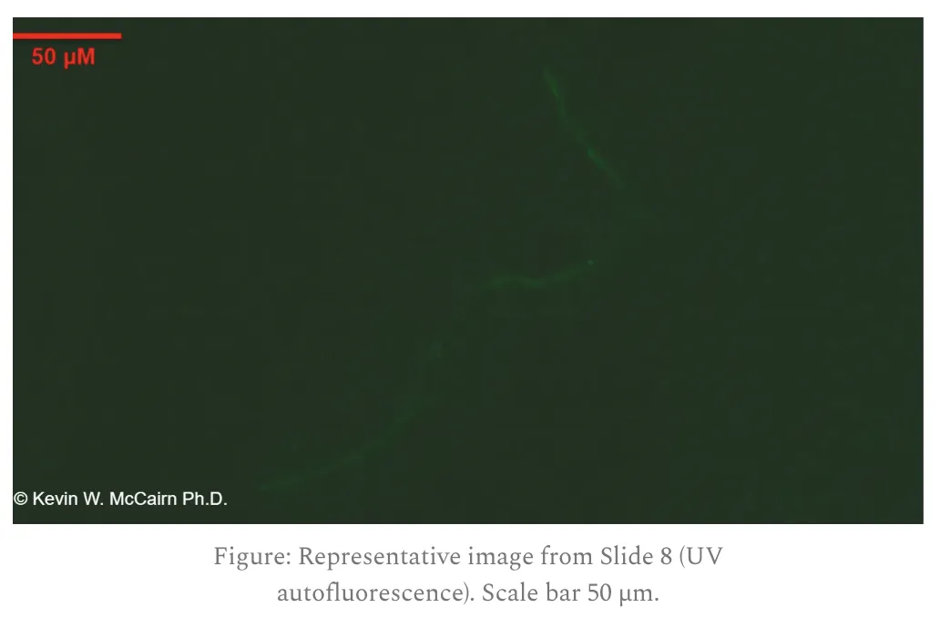
When McCairn asks his followers to send samples of blood to him on a slide in the mail, he supposedly finds fibrin clots with a diameter of about 10-30 µm in a large fraction of the samples. So if the density of clots per ml in his samples is representative of the overall density of the clots per ml in the bloodstream, then are there thousands of clots circulating in the bloodstream indefinitely, so that once the clots reach the arterial capillaries, they pass through the capillary bed to the veins, go back to the heart, and go back to arteries again? And if the clots are somehow induced by vaccination, then how do they remain in circulation even years after vaccination?
ChatGPT said that a clot with a diameter of 20 µm wouldn't pass
through the capillaries, which have a diameter of only about 5-10 µm:
[https://
No - a fibrin clot of the size you described (20 µm in diameter × 1 mm in length) could not "loop" repeatedly through the circulation. Here's why:
1. Size relative to vessels
Capillaries: ~5-10 µm in diameter.
Your clot: 20 µm in diameter, twice as wide as the largest capillaries.
Red blood cells deform to pass; fibrin clots cannot.
Thus, such a structure cannot traverse capillaries.
2. Artery-to-vein transfer
For blood to move from the arterial side to the venous side, it must pass through the capillary bed.
Since the clot is wider than capillaries, it would become trapped at the first capillary network it encounters.
It would not emerge on the venous side.
3. Circulatory "loops"
Only cells and small deformable particles (RBCs, WBCs, platelets) can continuously recirculate.
Solid emboli (clots, cholesterol crystals, etc.) either:
Lodge in a vessel (causing occlusion), or
Fragment into smaller pieces until they are small enough to pass further downstream.
The capillaries at the tip of a finger have a diameter of about 5-10
µm, so I was wondering if they wouldn't be too narrow for McCairn's
fibers to pass through in order for the fibers to end up in a finger
prick sample: [https://

McCairn instructs his followers to do the finger prick samples using a small instrument that is known as a lancet, which creates a puncture in the fingertip. However actually the lancet might reach deep enough to penetrate the arterioles and not only the capillaries, because ChatGPT said:
Capillaries in the fingertip are far smaller than a 20 µm clot:
Typical capillary lumen diameter: ~5-10 µm, often just wide enough for a single red blood cell (≈7-8 µm) to deform and pass through.
Precapillary arterioles / postcapillary venules: somewhat larger (≈10-30 µm), but true capillaries in the papillary dermis are in the 5-8 µm range.
Because of this:
A 20 µm fibrin clot would not be able to traverse capillaries; it is roughly 3-4× larger than the lumen.
However, if such a microclot were already present in a postcapillary venule or small arteriole in the dermal plexus (where diameters can be >20 µm), it could potentially be carried into the superficial circulation and then extravasated through a lancet puncture.
ChatGPT said that the arterioles start at a depth of about 0.3-0.6 mm beneath the surface of the fingertips:
In the fingertip, the vascular network is stratified. Based on microanatomical and histological studies of digital pulp and nail-bed skin:
Capillary loops: extend into dermal papillae immediately beneath the epidermis, beginning at ~50-150 µm below the skin surface.
Superficial dermal plexus: composed of arterioles and venules, located in the upper dermis at a depth of roughly 0.3-0.6 mm beneath the surface.
Deeper dermal/subpapillary arterioles (arising from the deep plexus): usually found at ~1-2 mm depth at the dermal-subcutaneous junction.
So, arterioles that feed the capillary loops near the fingertip start around 0.3-0.6 mm beneath the epidermis, while larger supply arterioles run deeper, closer to 1-2 mm.
And a standard finger prick lancet typically reaches a depth of about 1.5-2.0 mm. So I guess feasibly a blood clot might get stuck in arterioles because it can't pass through the arterial capillaries, and then it might get released if you do a finger prick that reaches the arterioles. So it wouldn't necessarily be anomalous if a clot is too big to pass through the capillaries, if in fact the clot was not circulating freely in blood, but it was stuck before the arterial capillaries in the fingertip.
But regardless, I consider it unlikely that the fibers McCairn finds in blood samples are blood clots, and so far McCairn has not even been able to show that his fibers are made of fibrin.
McCairn now also posted the image below, and he wrote: "I've found a more clear image of the linear clot, but
it's at a lower 4000X, the 5000x are all noisy. The reason for looking
at high mag is to dispel the notion that it's cotton which shows
pronounced linear striations. It's clear in this sample that the surface
is dimpled, non-striated, and has nodules consistent with other
amyloidogenic clots." [https://

The 4000x image shows a part near the end of the fiber towards the right side of the image (which was demonstrated by a second image in McCairn's tweet, but I didn't manage to save the second image before his tweet was marked as age-restricted content, and I don't have a phone so I'm not able to verify my age).
The new image shows a part close to the end that is about 9 µm wide, but the earlier 200x image showed a part closer to the middle that was about 13 µm wide.
ChatGPT said: "Cotton fibers from Gossypium appear flattened and twisted along their length because the central lumen collapses as the fiber matures and dries, giving the middle portion a ribbon-like, C-shaped cross-section. However, at the fiber ends, the lumen is less collapsed and the structure remains more rounded, so the ends typically appear smoother and more cylindrical under the microscope."
The earlier 200x image clearly showed a more flat and twisted shape:
[https://
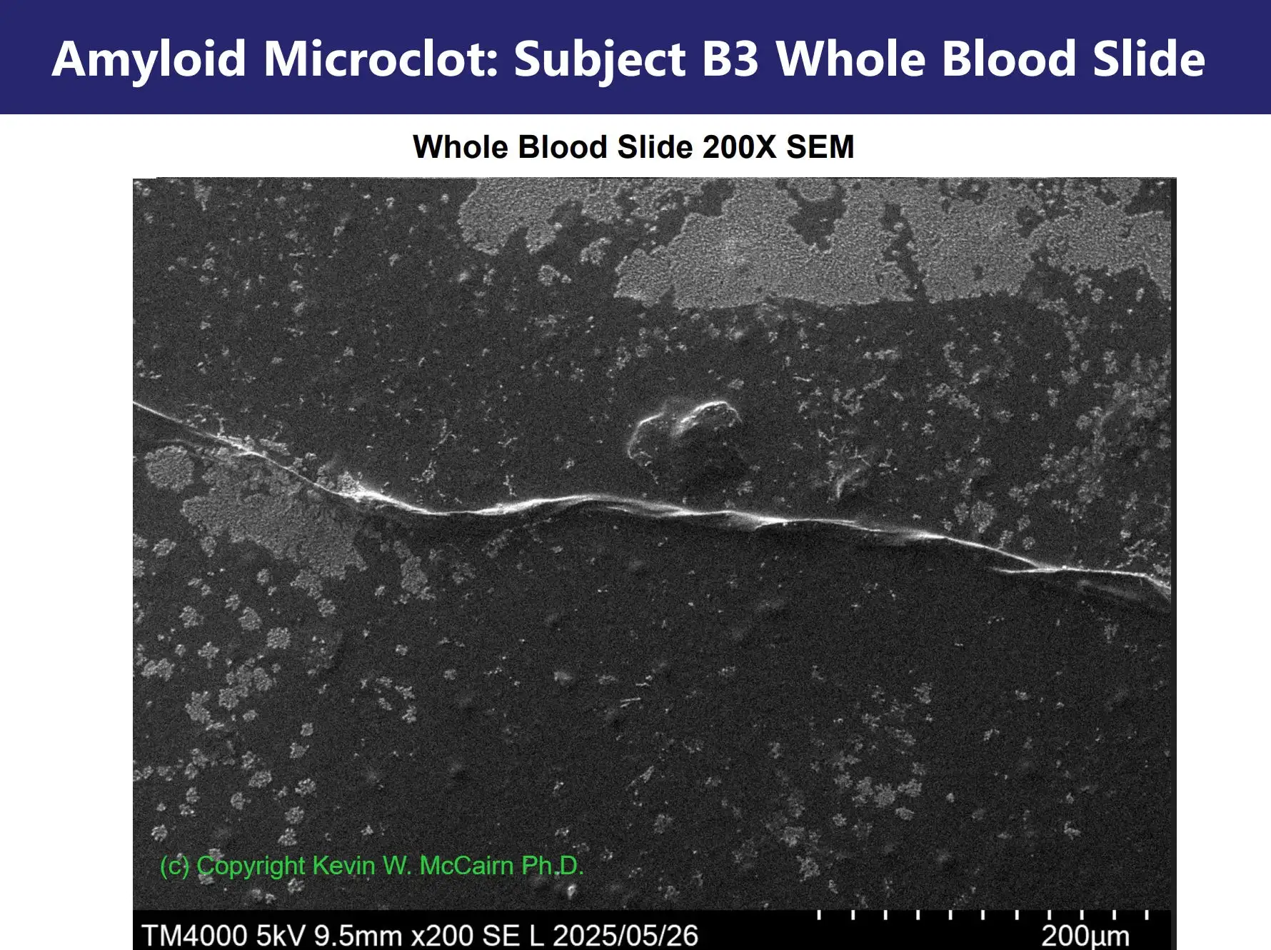
I told McCairn that his new image was so blurry that subtle striations might be hard to see. But then McCairn pointed out that the fiber also had "protruding nodule/nucleation sites", which I think meant the small ball-like specks that were attached to the fiber:

Then I posted a microscope image of fibers in dust with specks attached to them, and I asked McCairn if his "nodules" or "nulecation sites" were just specks of dust. But he said it was unlikely because his fiber had been soaked in buffer and dye:

I pointed out it would be easy to do an experiment where you took a sample of dust that contained fibers with specks attached, you soaked the sample in buffer and ThT, and checked if the specks remained adhered to the fibers. And I posted this response from ChatGPT:
Yes, dust specks could remain adhered to a microscopic fiber even if the fiber has been soaked in buffer and ThT, but the likelihood depends on several factors:
Nature of the Fiber Surface
- If the fiber is hydrophilic (e.g., cellulose or proteinaceous material), buffer and ThT will wet it well, which can loosen loosely attached dust particles.
- If it is hydrophobic (e.g., certain synthetic polymers), surface tension effects may cause dust specks to stay trapped.
Dust Particle Properties
- Dust is heterogeneous: it can include inorganic particles, organic matter, fibers, salts, and lipids. Some of these bind electrostatically or hydrophobically to surfaces and are not easily washed off.
- Small particles (<1 µm) may adhere strongly through van der Waals forces.
Buffer and ThT Effects
- Aqueous buffer itself usually does not solubilize dust (unless the dust contains salts).
- Thioflavin T (ThT) is a small, charged, aromatic dye. At the concentrations typically used, it does not act as a detergent, so it won't remove particles strongly bound to the fiber.
- ThT may even interact with hydrophobic dust or fibrous debris, leading to retention rather than removal.
Mechanical Handling
- Passive soaking is often insufficient to dislodge strongly adhered dust.
- Agitation, sonication, or filtration would be more effective.
McCairn told me I should get my own lab equipment to do the experiment. And in fact I have thought of buying some basic equipment so I could do experiments like this myself. But McCairn should be interested in performing the experiment himself, in case he relies on the specks attached to his fiber as evidence that the fiber is not made of cotton.
McCairn did a 3-minute video where he instructed his followers on how
to take a sample of blood from their finger and send it to him on a
glass slide. At the end of the video, he smears the droplets of blood so
that they're spread out over the glass, but the blood is left exposed to
open air, and he says "let these dry overnight in a
dust-free environment": [https://
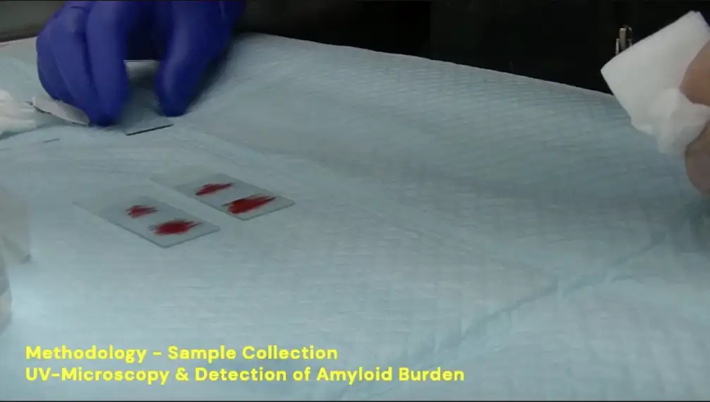
How many of his followers actually had a perfectly dust-free environment at their home? He didn't instruct his followers to put any cover over the slides so dust wouldn't fall on the slides. And he didn't instruct his followers to check afterwards that no fibers of dust had landed on the blood while it was drying.
Ian Musgrave is a molecular pharmacologist who has experience doing
electron microscopy of amyloids, but he said that McCairn's amyloid
fibril was a cellulose fiber: [https://
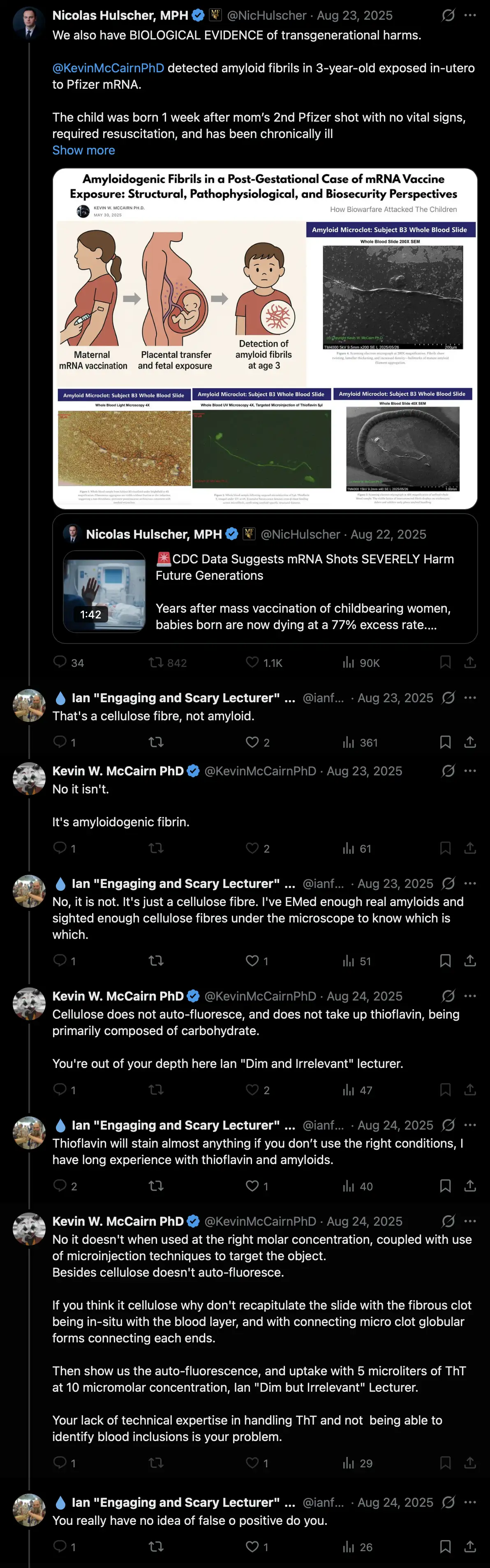
When McCairn said that cellulose doesn't autofluoresce, he forgot to mention that cellulose fibers can be strongly autofluorescent if they have been bleached white or washed with a brightening laundry detergent, like how white t-shirts are autofluorescent under UV light.
The Died Suddenly movie was produced by Lauren Witzke, who was Stew's
producer until 2024. In April 2024 Stew Peters tweeted: "The @DiedSuddenly_ account has been
hijacked by @LaurenWitzkeDE et al and is
currently not being run by the Stew Peters Network." [https://
In May 2025 the Died Suddenly crew announced that they were raising
funds for a new film called "Died Suddenly 2: Nano
Sapiens", which was going to be directed by Matthew Skow, who
directed the original Died Suddenly film together with Nicholas
Stumphauzer. [https://
Jikkyleaks said that the cast of the film was full of "glowies", but Kevin McCairn replied "LMFAO at the list of cretins in that list": [https://

One of the people featured in the film was Maria Zeee, but a few weeks earlier, McCairn went on Maria Zeee's show together with Richard Hirschman, who was the main star of the original Died Suddenly film:

Several other people in the Nano Sapiens film are also connected to
the story about the clots: [https://
Ana Maria Mihalcea: When she looked at
Hirschman's coffee ground clots under a microscope, and she presented a
video of blinking lights as evidence that the clots contained quantum
dot micro robots, Hirschman told her: "You got some
pretty daggum solid proof behind you when you're showing the actual
images, you're showing the blinking lights". [https://
La Quinta Columna: I have found no reference to
a new type of white fibrous clots before January 2022 when Jane Ruby
first broke the news about the clots. But Jane Ruby also says that she
broke La Quinta Columna's story about graphene oxide. [https://
Youngmi Lee and Daniel Broudy: Their paper about
nanotechnology in COVID vaccines was published by the journal IJVTPR.
IJVTPR has also published the only scientific paper about calamari clots
I have found, which was coauthored by Daniel Santiago and John Oller,
who are both members of the editorial board of IJVTPR. [https://
David Nixon: He is a member of NZDSOS, which
promoted An Unholy Triad, and which was co-founded by Matt Shelton who
was credited in An Unholy Triad. [https://
Steven Quay: Greg Harrison did a video where he
claimed that Quay's paper explained why the spike protein induced the
formation of calamari clots. [https://
In June 2025 Nicolas Hulscher posted a tweet where he promoted
McCairn's Substack post about the 3-year-old's fiber. Hulscher said the
fiber was an "amyloid fibril", even though
the diameter of the fiber was 3 orders of magnitude bigger than the
diameter of an amyloid fibril: [https://

Nicolas Hulscher is the "foundation administrator" of the McCullough Foundation, and he runs the Twitter account of McCullough Foundation. Peter McCullough is the Chief Scientific Officer of The Wellness Company.
In May 2025 McCairn and Hirschman went on Maria Zeee's show, which is now part of Vigilant News Network which was founded by TWC's co-founder Foster Coulson:
TWC's "chief marketing officer" used to be
Christopher Alexander, whose work experience includes having "successfully secured over 300 million dollars in
contracts for Information Operations, PSYOP, and intelligence
support" and being "recognized as a leader in
disinformation, misinformation, and counter-propaganda
campaigns". [https://
Accounts related to TWC seem to be artificially amplified by bots on
Twitter. In 2024 I scraped the reposts of about 80 accounts that I
suspected to be bots that promoted content about COVID from the
controlled alternative media. When I sorted accounts by the number of
reposts by the suspected bots divided by the number of followers, and I
excluded accounts that were not related to COVID and accounts that had a
low number of followers, McCullough Foundation ranked 2nd highest. [bot2.
Makis promotes ivermectin and fenbendazole as miracle cures for turbo
cancer, but before COVID both of them were presented as cures for
Morgellons disease: [https://

A forum post from 2016 said: "I've also read that
a lot of people who are suffering with morgellons have had some huge
success with fenbendazole." [https://
In 2024 Hulscher and McCullough coauthored a paper with Raphael
Stricker, who is possibly the main person responsible for popularizing
the Morgellons disease hoax. Stricker also coauthored a paper in 2021
with McCullough and Harvey Risch, who are both members of the "Chief Medical Board" of TWC: [https://

The term Morgellons disease was coined by Mary Leitao. The earliest
paper about Morgellons disease that is structured like a scholarly paper
is probably a paper from 2006 that was coauthored by Virginia Savely,
Mary Leitao, and Raphael Stricker. [https://

In 2019 the California Medical Board issued a complaint against
Stricker because he prescribed ivermectin as a treatment for Morgellons
disease without obtaining informed consent. [https://
In a video that Ana Mihalcea did with Hirschman, she said that
calamari clots are "made from these filaments that
Clifford Carnicom calls cross-domain bacteria or Morgellons
filaments", and she showed these images of Morgellons filaments
that she found in Hirschman's clots: [https://

Then Hirschman told Mihalcea "I had a live blood analysis of my own blood and my blood had some of those filaments in it too" (even though it's not clear if by the filaments he meant Morgellons filaments or some other type of filaments). [55:26] As evidence that Hirschman's clots contained quantum dot microrobots, Mihalcea showed a microscope video of blinking lights, but Hirschman told her "you got some pretty daggum solid proof behind you, when you're showing the actual images, you're showing the blinking lights". [1:00:57]
The only scientific paper about calamari clots I have found was
published in the journal IJVTPR (if I employ a very
loose definition of a scientific paper that includes pseudo-scientific
papers masquerading as scientific papers). One of the people on
the editorial board of the journal is Shimon Yanowitz. [https://

The term "turbo cancer" was first
introduced in September 2021 in Arne Burkhardt's pathology conference.
[turbo.
In reality the fibers in the image above have several characteristics of cotton, because they are about 2 to 3 blood cells wide, they have a flat body with a cupped C-like shape, and they have the kind of lengthwise twists that are called convolutions in fibers of cotton.
One of the people in McCairn's circle is Johanna Deinert, who said
she advised Arne Burkhardt on immunohistochemistry, and I believe she
even helped run his website, which is rather suspicious considering how
Burkhardt appears to have been a Quinta Columna type plant in alt media.
[https://
Added later: Apparently Mary Leitao also claimed that Morgellons
filaments were autofluorescent: [https://
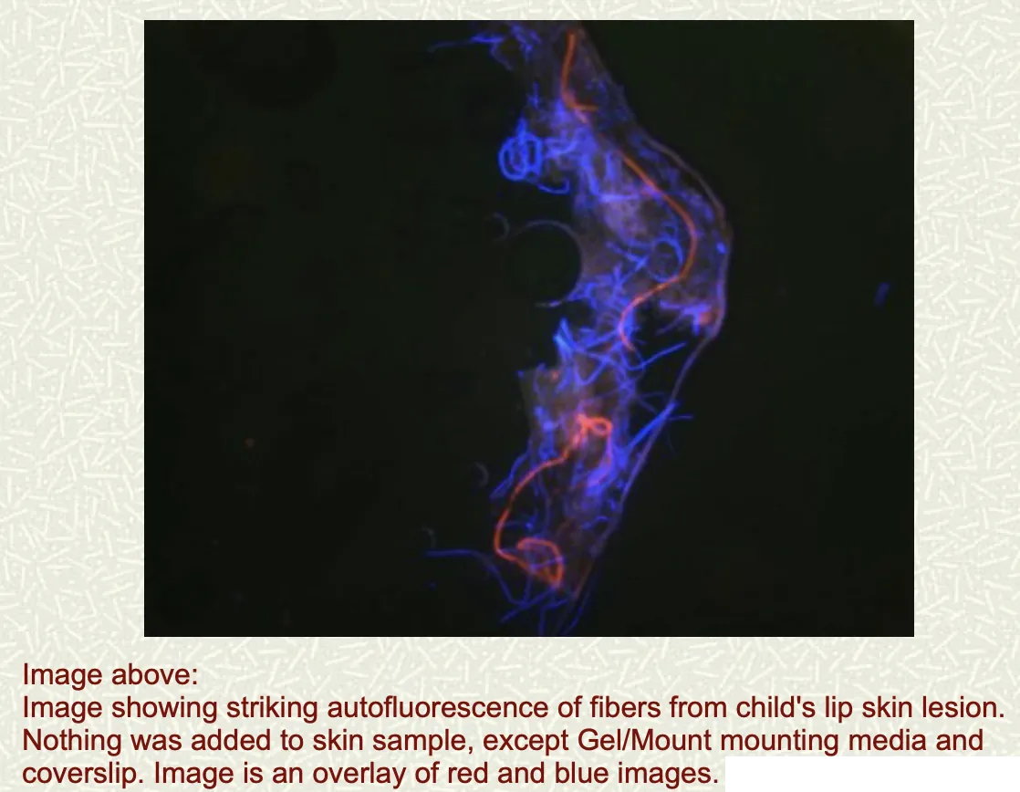
Adam Finnegan promoted this video McCairn did with Nicolas Hulscher,
where McCairn presented his neo-Morgellons theory about how vaccinated
people have developed a scary new pathology which causes their body to
grow fibers that look like textile fibers: [https://
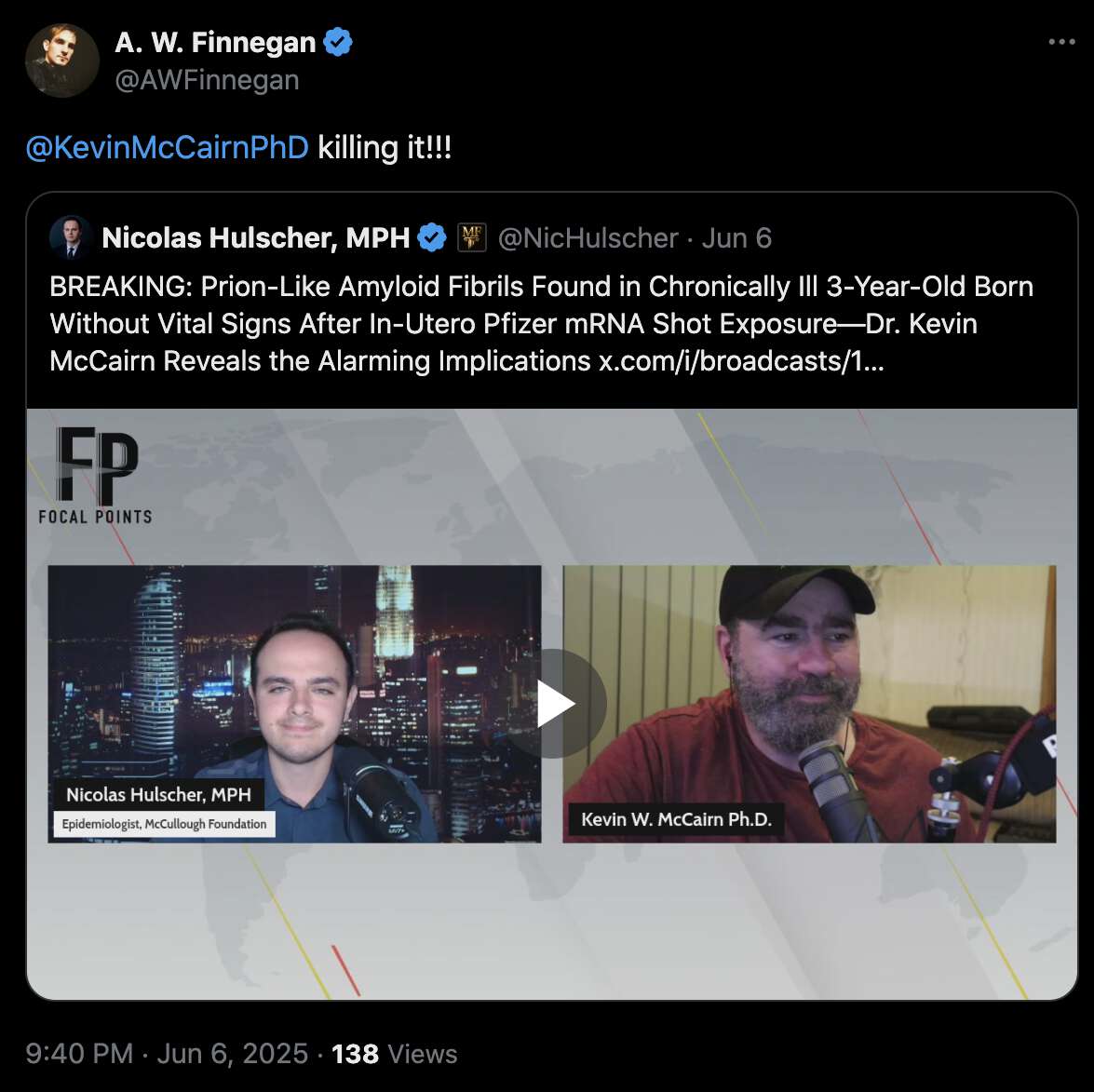
Kevin McCairn has done multiple videos with Adam Finnegan, and Finnegan is on McCairn's Discord and he is promoted by many people in McCairn's circle.
In 2021 Finnegan wrote an article about Morgellons disease, where he
claimed that he had witnessed Morgellons disease in his extended family
members, he characterized the Carnicom Institute as a credible source,
and he suggested that the presence of microplastics in rain might mean
that Morgellons is spread through chemtrails. He wrote that Morgellons
disease was the result of "adverse events in
nanotechnological failure", and he compared Morgellons filaments
to nanowires produced by genetically modified viruses (even though the fibers that are said to be Morgellons
filaments are visible with an optical microscope, but nanowires are
not). [https://
Finnegan also showed a photo of plastic fibers in rainwater, and he
compared it to Mary Leitao's photo of Morgellons filaments: [https://
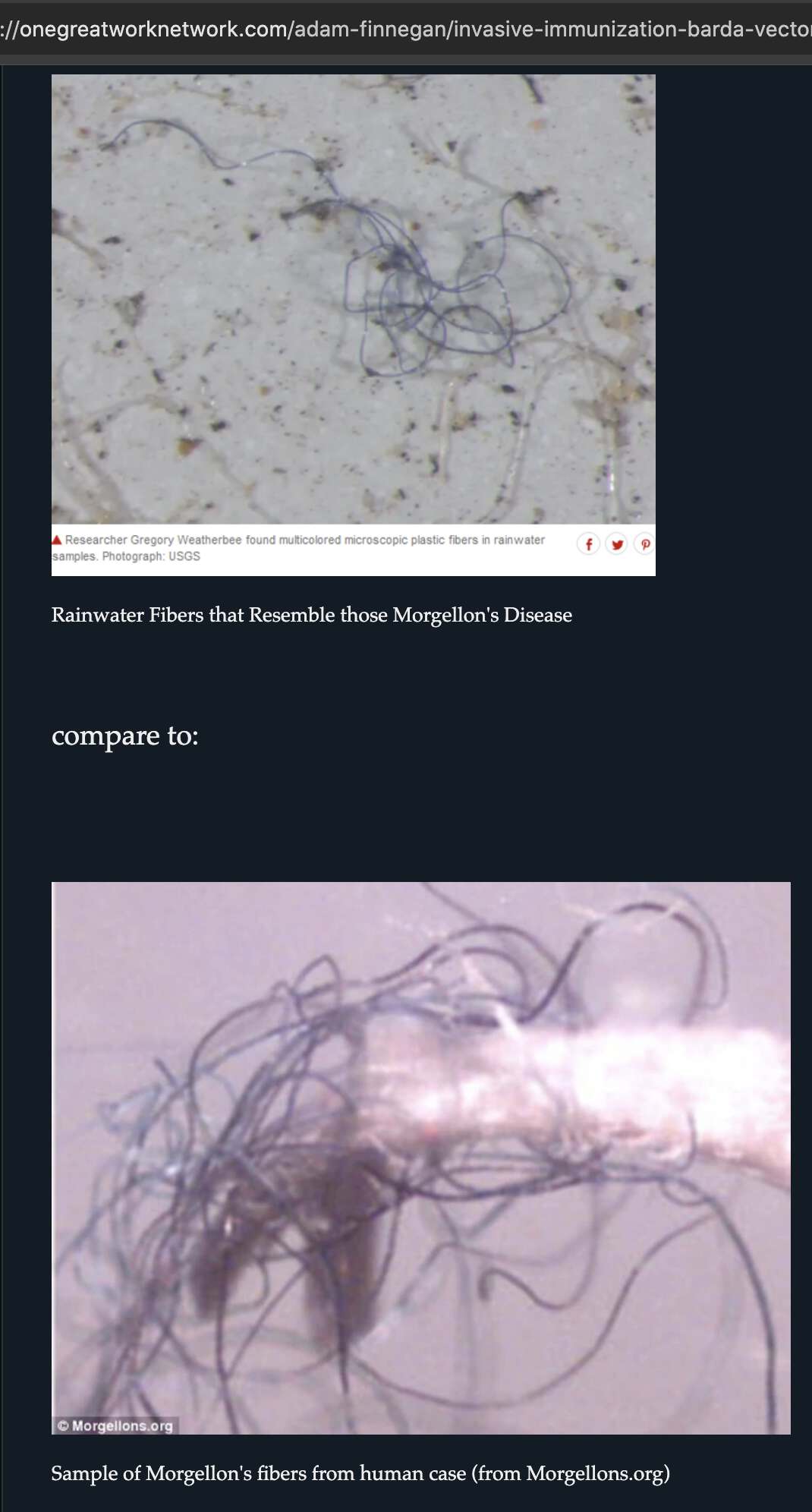
Finnegan is a regular guest on the channel of the son of a high-level
FBI agent, who also recorded an audio narration of Finnegan's book, and
who appears to be Finnegan's biggest advocate in alt media. The son of
the FBI agent launched his YouTube channel together with a Jew who said
that he had homies at French Mossad, and that he worked with Dutch
intelligence in New York: [https://

Edited in 2026: The rest of this section has been split off into a
dedicated article about Finnegan and his mentor John Loftus:
lyme.
A Twitter user called Tom Czerniawski said that prions from vaccines
will kill so many people that all human life on earth will end, and he
said it was not hyperbole or exaggeration. Kevin McCairn replied to him
suggesting that the prions will result in an evolutionary bottleneck
event: [https://
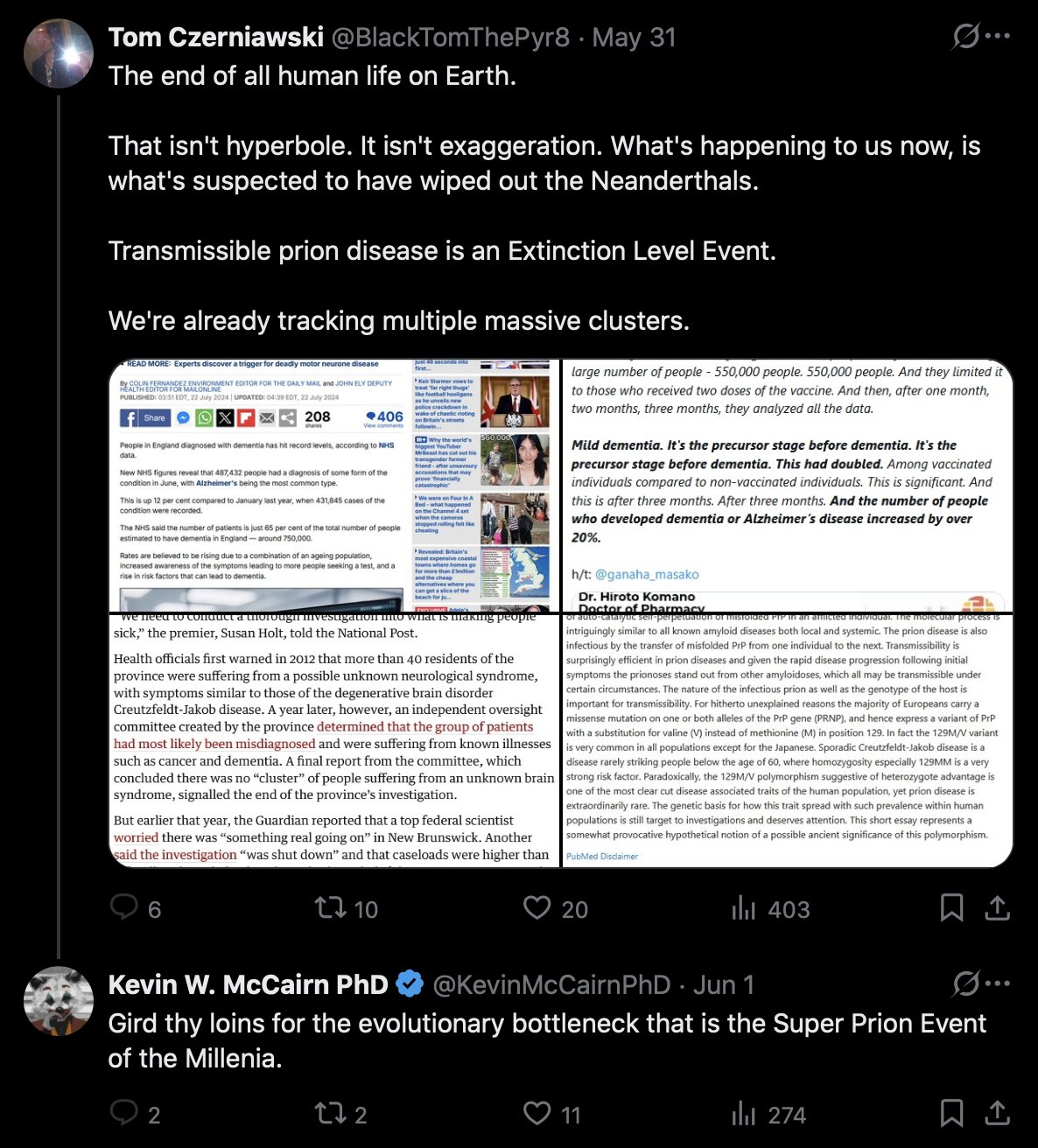
Earlier Tom Czerniawski was going around asking billionaires to
donate money to McCairn, because he said that if McCairn receives a few
hundred thousand USD in donations, he could "SAVE
EVERYONE FROM DYING HORRIBLY": [https://
Here Tom said that the clots will cause a cataclysmic pole shift to
arrive more quickly, and Ethical Skeptic agreed with him: [https://

One time when I told McCairn that his followers have the same kind of
cult mentality as no-virus people, he said "no they
don't" and "it's not like that at
all". [https://
Added later: As further evidence that McCairn's followers are members
of an apocalyptic cult, the pinned tweet of one of his fans says that
the spike protein has a prion epitope that is creating an
extinction-level super prion event (which somehow
only McCairn has been able to reveal through his scientific
genius): [https://
Added in 2026: Now Tom suggested that McCairn was an angel of
salvation, who was sent to earth by divine providence to save humanity:
[https://
Here Tom seems to have implied that people like McCairn are the best
humans on the earth: [https://

Black Tom also said that Jikkyleaks has saved countless millions of
lives, or potentially billions down the road: [https://
In May 2025, Kevin McCairn published a Substack post about how he supposedly found an amyloid fibril in the blood of a 3-year-old boy. His story went viral after Nicolas Hulscher published a Substack post about the story. Hulscher's post was then copied by Frank Bergman, who is the most prolific author on the fake news site Slay News. Bergman's article was promoted by the Died Suddenly account on Twitter, which posted a screenshot of Bergman's article copied to another fake news site called TRUTH11.COM:

A few days later, a paper about how vaccinated people were magnetic was published in the Indian predatory journal IJIRMS, which has also published papers by Hulscher and McCullough. At first the paper didn't get too much attention, but a few days later it went viral after Hulscher wrote a Substack post about the paper, and then Hulscher's article was again copied by Frank Bergman, and Bergman's article was promoted by Died Suddenly:

Nic Hulscher also posted similar AI-generated infographics promoting
both stories: [https://
Frank Bergman is likely a fake person, and his portrait picture
appears to have been generated by thispersondoesnotexist.com or by some
other interface to StyleGAN: [https://

Slay News is run by British people from a company called Evil Corp Ltd. They also run another fake news site called News Addicts, where the most prolific author is Hunter Fielding who has an AI-generated portrait picture. Articles by Hunter Fielding are also published on the British fake news site Expose News.
There's a British Substack author who uses the pseudonym Iggy Semz, who has been a guest on McCairn's stream. After I started compiling evidence that the clots were fake on McCairn's Discord server, Iggy launched a massive character assassination campaign against me, and he flooded the server for days with posts where he attacked me. His profile picture appears to have been generated by thispersondoesnotexist.com, because when I opened the full version of his profile image on Discord, it had a black rectangle at the same spot where thispersondoesnotexist.com adds a watermark, and the eyes in his picture are located at the same spot as the eyes in Frank Bergman's picture:
The lead author of the magnetism paper was Pasi Suominen who is a
member of Jikky's mouse army, so Jikkyleaks said that if the paper "got through peer review then it's got merit" (even though the paper was published in an Indian
predatory journal): [https://
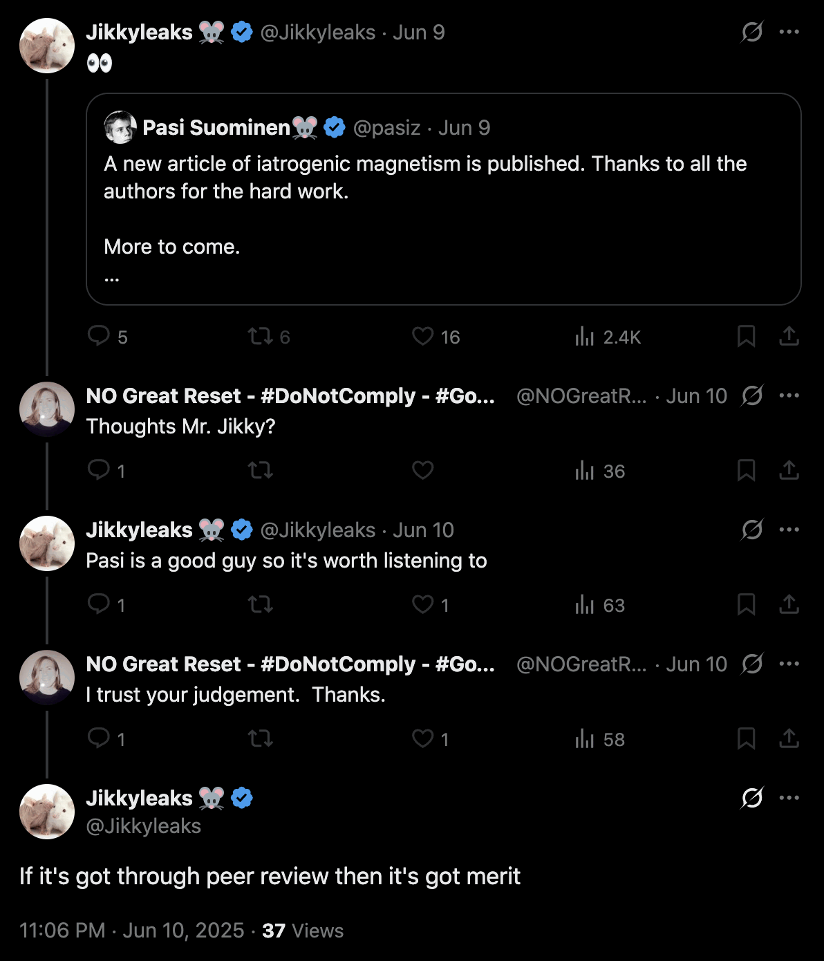
I'm calling Pasi Suominen's group the Finno Columna, because the
story that graphene oxide made vaccinated people magnetic was originated
by La Quinta Columna. Quinta Columna came up with #MagnetGate and
#BluetoothGate before Jikky came up with his first gate: [https://
A Twitter user called Jesus4AllAlways posted the images below and
wrote: [https://
My family of 5 are unlucky patients who took pFIZER Feb 2021 double jab In Rockaway NJ.
Using an Olympus CX 43 lab microscope from January 3, 2024 regular for some reason in November 2024 I observed the fiber looking structures in my blood. I began to research what they were.
Suspicious of the PJab in January to May 2025, I checked my wife and children. Bad news.
I observed the same strange structures in each of them.Pic 1 shows my May 25 observation of myself
Pic 2 is 22 year old child
Pic 3 is wife
Pic 4 is 20 year old
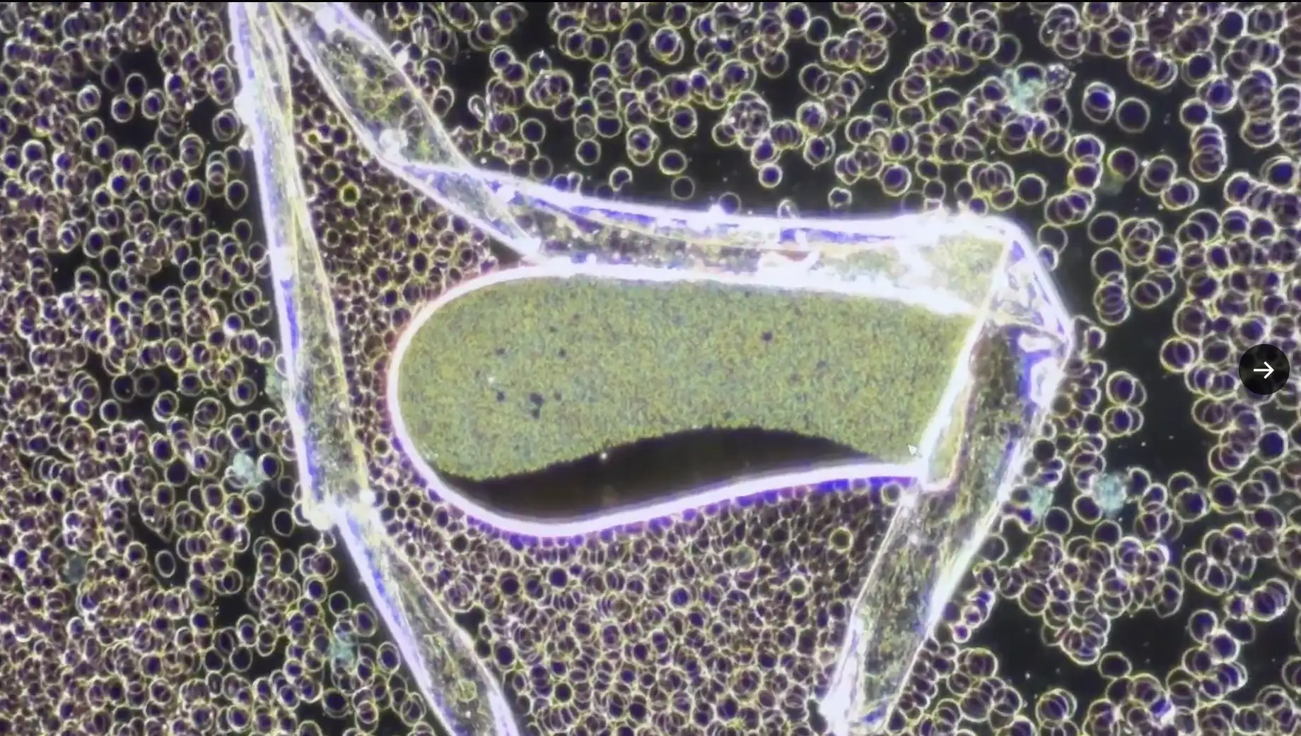
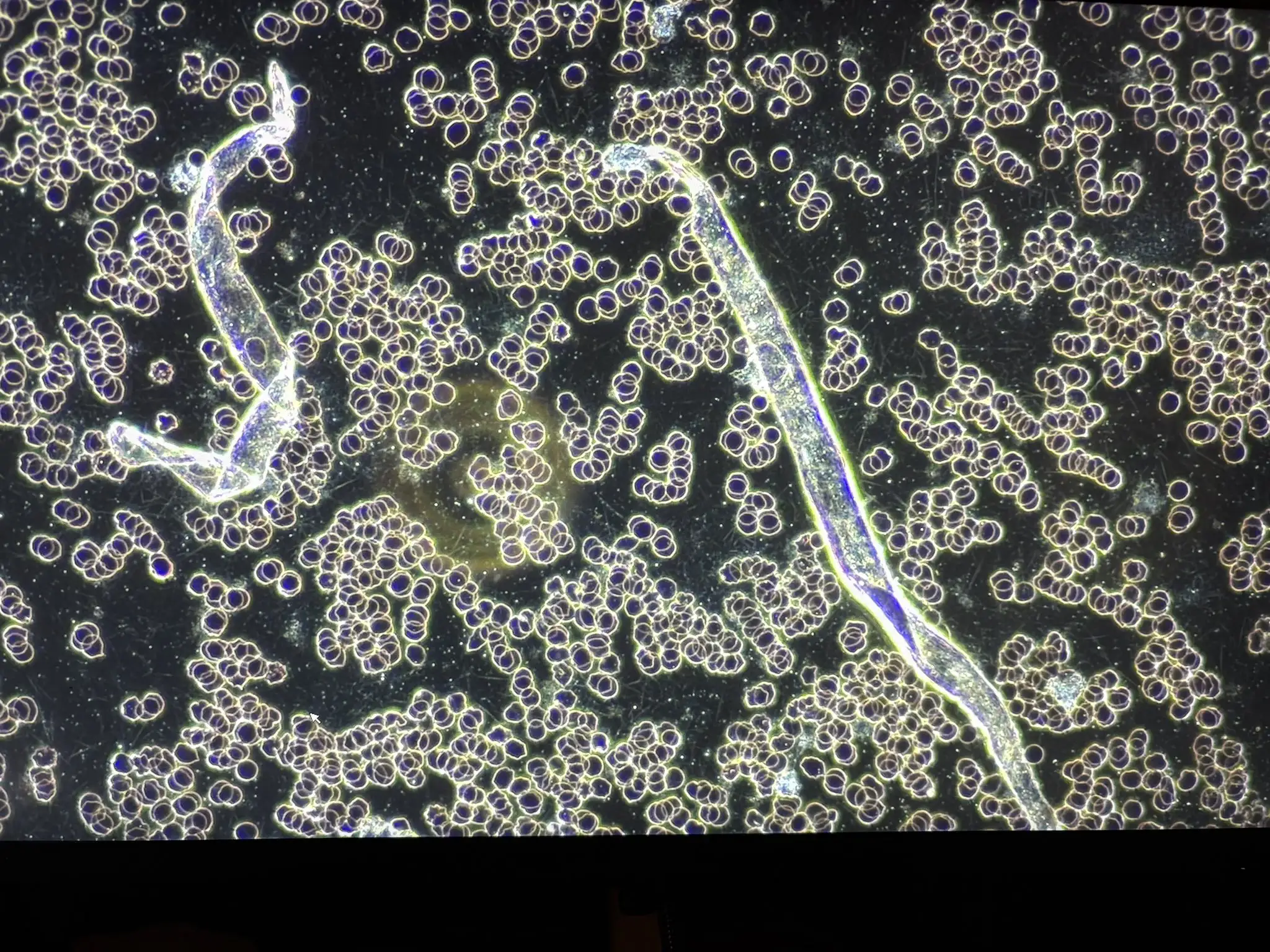
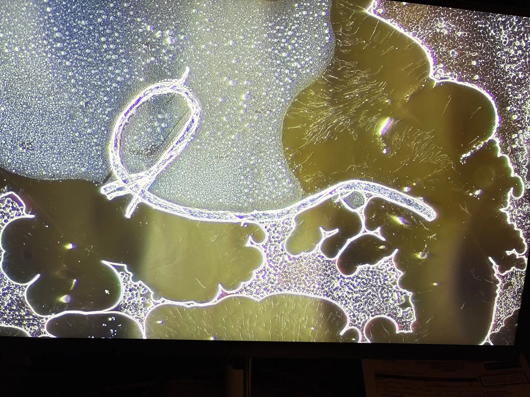
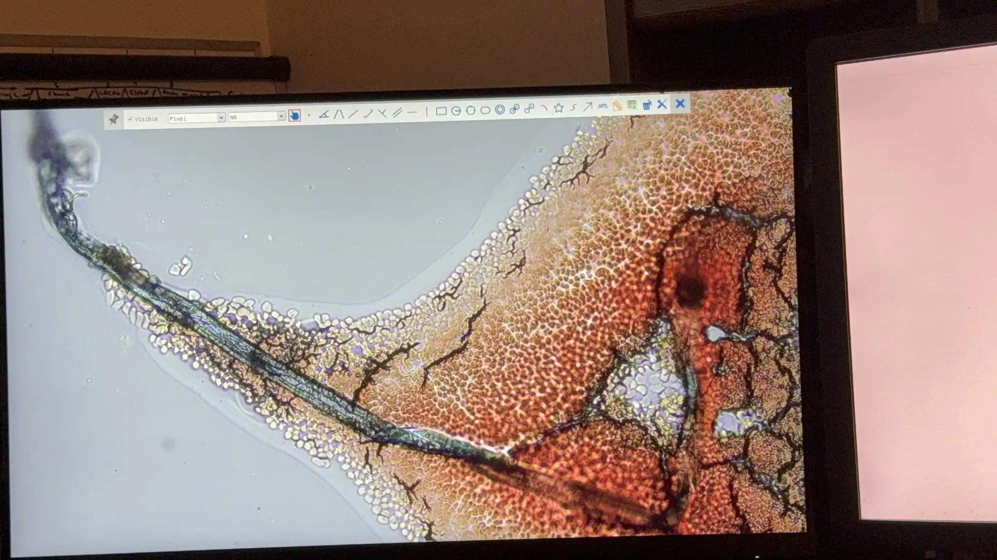
(The first image was actually a video clip, but I included a screenshot of the video above. In the video Jesus4AllAlways said that the cells were white blood cells, even though they're actually red blood cells.)
In a reply to Jesus4AllAlways, McCairn suggested that one or more of
the structures in the images were "amyloidogenic
fibrin": [https://
The structures in the first two images are about 3-4 times wider than the red blood cells, but red blood cells have a diameter of about 6-8 µm, so the diameter of the structures is roughly consistent with the fibers McCairn has found in blood samples, which have had a diameter of about 10-30 µm.
Some of McCairn's fibers have been flat like the structures in the first two images, but his other fibers haven't looked similarly flat. But even one of McCairn's flat fibers had a lengthwise fold running along the middle, which was missing from the structures in the images by Jesus4AllAlways. And the fibers in McCairn's microscope images haven't looked as translucent as the structures in the first two images. And McCairn's fibers haven't had the kind of folds of about 90 degrees as in the first two images, where you can see different parts of the folded structure overlapping each other. And Jesus4AllAlways didn't even do UV microscopy or ThT staining.
So then why was McCairn ready to declare that the structures in the images were "amyloidogenic fibrin"?
In 2022 when Shimon Yanowitz was on Maria Zeee's show, he showed an
image of the structure below, which has similar folds as the ribbons in
the blood of Jesus4AllAlways. Yanowitz said the structure was a killer
tube which could be programmed to kill the host, and he said: "There are now billions of people walking around with some
structures inside of them that could be commanded to open up and release
the payload upon external command at will." [https://

This is what ChatGPT said about Shimon's killer tube:
This image shows a ribbon-like structure under a microscope, viewed with polarized light or differential interference contrast (DIC), based on the high-contrast, crystalline-like edges. The object in question is highly consistent with a synthetic microplastic fiber, most likely polyester, nylon, or another textile-derived filament.
Characteristics Supporting This:
Flat, crumpled ribbon appearance typical of synthetic fibers.
High birefringence (bright glow under polarized light), which is characteristic of plastics.
No internal biological structure (e.g., no nuclei, organelles, or cellular components).
The irregular folding and twisting are typical of how synthetic fibers behave when dried or compressed.
Ana Mihalcea said that similar translucent ribbons with folds were
"hydrogel filaments": [https://

ChatGPT said that Mihalcea's hydrogel filaments were likely to be synthetic fibers:
Left Image:
The background consists of what look like red blood cells (RBCs) - biconcave discs, tightly packed.
The ribbon runs diagonally and has a smooth, semi-transparent appearance with light-scattering properties, suggesting it is not biological tissue.
The ribbon's uniform thickness and reflective surface suggest it could be a polymer fiber, perhaps from clothing or lab materials (e.g., polyester or nylon).
Right Image:
A similar structure is seen again, this time bent at an angle.
The bright refraction and internal stress patterns visible under polarized or brightfield illumination are typical of synthetic materials, not biological ones.
The presence of RBCs again near the ribbon supports that this is a foreign object in a biological sample.
Conclusion:
The ribbon in both images is most likely a synthetic fiber accidentally introduced into the sample - possibly from clothing, gloves, wipes, or sample coverslips. It is not a biological structure.
Similarly when I asked what the ribbon in the video by Jesus4AllAlways was, ChatGPT answered: "The structure has sharp folds and flat surfaces, consistent with plastic sheeting or synthetic ribbon fibers, not biological tissue."
Added later: I asked McCairn if he still stuck to his claim that the
images by Jesus4AllAlways showed "amyloidogenic
fibrin", and if by "amyloidogenic
fibrin" he referred to the flat ribbons with folds. He indicated
that he did refer to the flat ribbons with folds, and he said: "I would say from that image of that the ribbon like
structure is consistent with the morphology of amyloid fibrillar forms
seen in long covid and vax injured." [https://
However I again uploaded Mihalcea's images of hydrogel filaments to ChatGPT, and I asked if the folds and flat shape were consistent with a fibrin clot. But ChatGPT said:
No - sharp folds like the ones in your image (especially the ~90° bend in the right-hand picture) are not characteristic of natural fibrin clots.
Here's why:
Fibrin mechanics: Natural fibrin fibers are semi-flexible biopolymers. When bent, they curve smoothly because the protofibril bundles resist sharp angles - you get arcs, not crisp corners.
Amyloid fibrin: Even when fibrin is converted into amyloid-like β-sheet-rich forms, it becomes stiffer and more brittle, but it still tends to fracture or kink irregularly rather than form a neat, flat, ribbon-like corner.
Your sample: The flat cross-section, uniform thickness, and clean angular bend are far more consistent with synthetic fibers (e.g., textile microfibers), which can hold a crease from manufacturing or folding.
In short, a true natural fibrin clot won't spontaneously develop a paper-fold-style corner under physiological conditions or during slide prep - unless it's being mechanically crushed between coverslip and slide, in which case it fractures rather than bends smoothly at 90°.
In July 2021 Philippe van Welbergen presented the microscope images
of vaccinated blood below, and he suggested that the translucent ribbon
with folds was made of graphene oxide. The ribbon is about 3-4 red blood
cells wide, so it has a similar diameter as the ribbons shown by
Jesus4AllAlways and Mihalcea: [https://
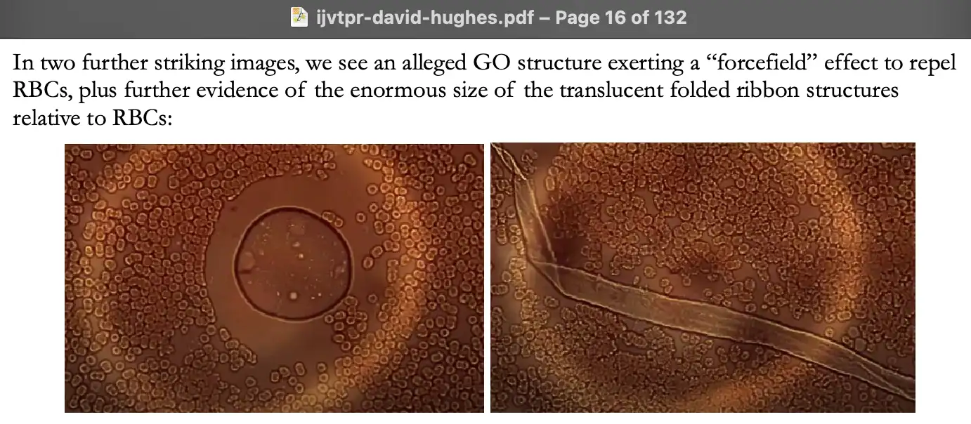
Just like McCairn, van Welbergen also suggested that the presence of ribbons was somehow a characteristic of unhealthy vaccinated blood:
In 2021 Jane Ruby did a video where she showed the photos below, and
she said: "Dr. Welbergen calls those gold structures
tubes, when he magnified them even further on a regular microscope. They
are actually in a tube form, and you can see the opening on either end
of those. Remember, this looks strikingly like the graphene oxide that
we saw under the regular microscope, from the Spanish researchers like
the Quinta Columna, where you saw that folded over protein that looked
like it was like a piece of Kleenex under a pane. And that's what this
is." [https://
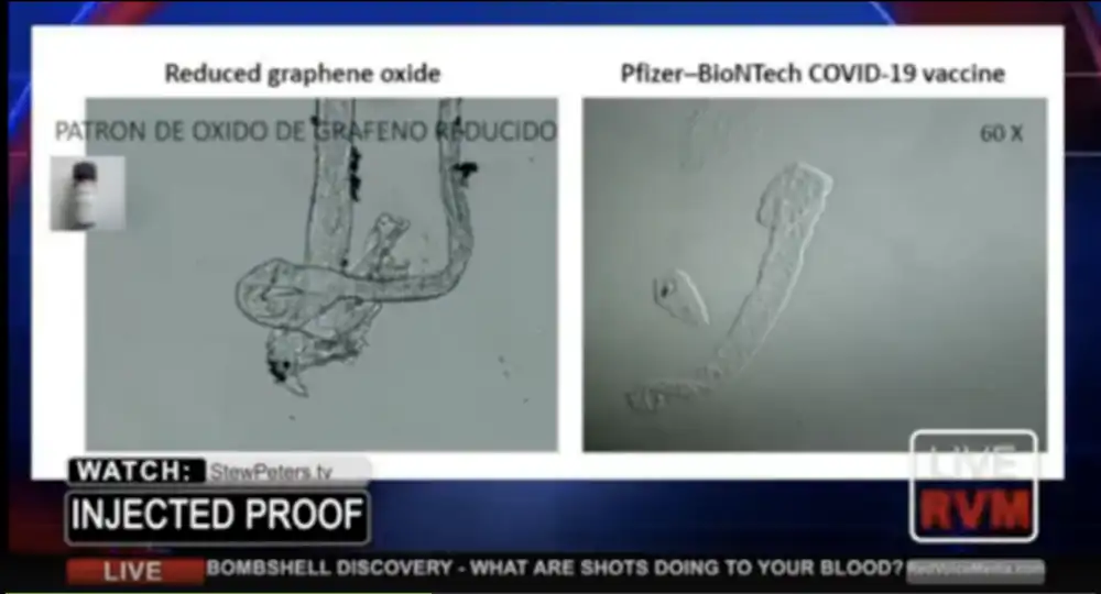
Kevin McCairn told me that the calamari clots are "very clearly blood vessel occlusions, composed of
amyloidogenic protein, that have the same properties as amyloidogenic
micro clots, that have been proven human tissue through genomic
testing". [https://
However I told him: "A clump of fibrin is not tissue. If you dipped some rubber in blood and tested it genetically, you would find human DNA from the blood, but that doesn't mean the rubber would be human tissue. It's possible the clots have a human origin, but I think it has not yet been proven." And I showed him this response from ChatGPT:
Calling such clots "human tissue" would be misleading. Here is why:
Blood clot composition: A fibrin-rich clot with little entrapped blood cells is still just a coagulum of protein (fibrin) rather than a structured tissue. It lacks organized cells, extracellular matrix, vasculature, or architecture typical of tissue.
Definition of tissue (in biology/medicine): Tissue is a collection of cells (of one or more types) and their extracellular products, organized to perform a function. Examples: epithelial tissue, muscle tissue, connective tissue.
Clot vs. tissue: A clot is an acellular polymer network (fibrin mesh) that sometimes traps platelets, leukocytes, and red blood cells. While it originates from blood (a fluid connective tissue), a fibrin clot by itself is not equivalent to "human tissue."
Terminology in practice: Pathologists or hematologists would usually describe this as "fibrin clot," "fibrinous exudate," or "thrombus" (if formed in vivo), but not "tissue."
McCairn posted an AI response which said that the clots are not human
tissue, but the clots can be still viewed as a "quasi-tissue scaffold", or the clots can be
considered to "function as novel pathological
pseudo-tissue": [https://

But I told him that those were bad copes, and that he is a quasi-scientist and he functions as a pseudo-scientist. He cannot even tell protein apart from tissue, or a peptide apart from a protein, or a micrometer apart from a micromolar, or a textile fiber apart from an amyloid fibril.
I told McCairn I hadn't found evidence that string-like fibrin clots
with a diameter of about 10-30 µm would be circulating freely in human
blood. He responded by showing the images below from a paper where the
senior author was Resia Pretorius, and he wrote: "Row B is a string like microclot that would be well over
100 micrometers, as it fills the whole micrograph, from whole blood, and
stained with ThT." [https://

I pointed out that the string-like segments on row B had a diameter of only about 1 µm. They also seem to be part of a web-like structure and not lone isolated fibers.
Then I asked ChatGPT to list the diameters of different types of fibers that appear as contaminants on microscope slides. Most types of fibers it listed have a diameter around the range of 10-50 µm, which matches the diameter of the fibers that McCairn has found in blood samples:
Synthetic Fibers
- Nylon: ~10-50 µm
- Polyester (PET): ~10-30 µm
- Acrylic: ~10-25 µm
- Polypropylene: ~20-50 µm
- Rayon (regenerated cellulose): ~12-20 µm
Natural Fibers
- Cotton: ~10-40 µm (flat, twisted ribbon-like appearance)
- Wool: ~15-40 µm (scaly surface)
- Silk: ~10-15 µm (smooth, uniform diameter, triangular cross-section)
- Linen (flax): ~12-16 µm
Mineral and Glass Fibers
- Asbestos: ~0.02-3 µm (often below light microscopy resolution, needle-like)
- Glass fibers: ~1-20 µm (uniform, cylindrical)
- Rock wool / slag wool: ~2-10 µm
Other Microscopic Fibers
- Cellulose fibers (paper fragments, wood pulp): ~10-50 µm
- Carbon fibers: ~5-10 µm
For example these cotton fibers are about 20 µm wide at the widest
point: [https://

In this image, the cotton and linen fibers both have a diameter of
about 20 µm: [https://
The next photo supposedly shows hydrogel filaments that Mihalcea
found in the blood of a corpse embalmed by Hirschman. The filaments are
about 2-3 red blood cells wide, which is about 15-20 µm, because red
blood cells typically have a diameter of about 6-8 µm: [https://

This fiber that Mihalcea claimed was a carbon nanotube also had a
diameter of about 2-3 red blood cells: [https://
These fibers in the blood of the son of Jesus4AllAlways are also
about 2-3 red blood cells wide: [https://
McCairn said that the fiber below was a "fibrillar form clot" that he found in his own
blood. The fiber is about 3-4 red blood cells wide at the widest point.
McCairn said the scale bar was wrong and it was supposed to be 50 µm
wide, but based on the size of the red blood cells, the scale bar might
be closer to about 100 µm wide: [https://
McCairn supposedly found the fiber below in the blood of Lyndsey
House in 2024. If the scale bar is accurate, then the fiber is about 15
µm wide: [https://

Robert Young also found a scary fiber with a diameter of about 3 red
blood cells in vaccinated blood: [https://
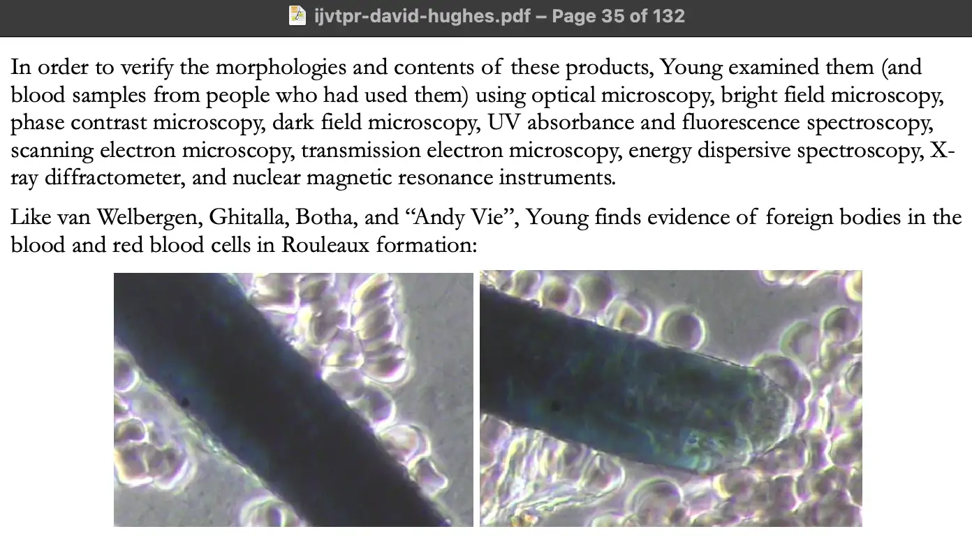
Clifford Carnicom's website has an archive of articles going back to
1999. His early articles focused on topics like chemtrails and HAARP,
but his first article about Morgellons disease is from 2006. [https://

However Carnicom failed to mention that both the diameter and the twisted shape were characteristic of a fiber of cotton. He also posted these images of fibers in the sample, which look like cotton because they have a flat cupped shape, and they have the kind of twists that are called "convolutions" in cotton:


In Carnicom's second post about Morgellons disease, he wrote about a
sample he is supposed to have received from another Morgellons patient.
He posted this close-up photo of one of the fibers in the sample, where
you can now see the flat cupped shape even more clearly: [https://

Carnicom wrote that the diameter of the fiber was approximately 40
µm, which would be about 3-4 times wider than his earlier fiber that he
said was about 10-12 µm wide. Fibers of cotton typically have a diameter
of about 10-40 µm, or sometimes a narrower range of variation is given
as 12-25 µm. However Carnicom's estimates of the diameter might have a
fairly wide margin of error, because in another post he estimated that a
structure in his image had a diameter between 3 and 10 µm, so his
maximum estimate was more than 3 times higher than his minimum estimate.
[https://
Carnicom said that his Morgellons filaments were composed of multiple
"sub-micron filaments" which were packed
together within a "bounding filament". By the
"sub-micron filaments", he might have
referred to segments of the fiber that were separated by the lengthwise
striations which are characteristic of cotton. In one post he wrote:
"The diameter of the bounding filament generally
ranges from 20-40 microns in thickness." [https://
McCairn has now started giving expensive treatment to people which supposedly removes the fibers from their blood that he initially called "amyloid fibrils", but that he started calling "fibrillar formations" after I told him he was misusing the word fibril. Now he seems to be calling them "aggregates" instead.
Earlier McCairn gave the impression that the fibers he found in blood
samples were circulating freely in the bloodstream, but me and another
user on his Discord pointed out that his fibers had a wider diameter
than capillaries, so the fibers wouldn't be able to pass through the
capillary bed. But then later I figured out that in case the fibers were
lodged in the arterioles before the capillaries in the fingertips, the
fibers might get released from the arterioles if you do a finger prick
sample, because typically a finger prick lancet would reach deep enough
to penetrate the arterioles and not only the arterial capillaries.
However it's a hypothesis I came up with on my own and that McCairn
didn't suggest to me (and I came up with the
hypothesis because I was trying to play along with the scenario where
the fibers that look like textile fibers would actually be clots made of
fibrin, which I was nearly certain was not true): [https://
Earlier I asked McCairn several times if the reason why his supposed
clots had a string-like shape was because the clots formed in a small
blood vessel and then got dislodged, McCairn came up with explanations
like that "there is a coherency in the
epistemological grounding of the amyloid PRION formation". [https://
However now when he did a video with one of his first patients,
McCairn said himself that the so-called "aggregates" are "lodged into
the entrance and exits of capillary beds", where he even used the
same verb "lodged" that I had earlier used
many times to refer to the hypothetical clots that are stuck before the
capillaries. He said: "So, what's the one thing that
we can point at right now, which is quantitative in a ways that the
aggregations in the plasma disappear? And, are they generated de-novo,
or are they a consequence of initial exposure, and then just sort of
building up over time? I'm still unsure. But what I can say is between
the DFPP - PA session, sorry - you were in the second treatment
protocol, in the pre-plasma - there was amyloid-like burden in the
pre-plasma that has gone in the post. So my thinking is that there's a
bulk movement of the plasma during filtration, and that's gonna drag a
whole bunch of these aggregates with them. It's coming back in clean.
And then if there's more that are sort of hanging on to the vasculature,
they're gonna then be washed off into the plasma again, which is why
we're seeing the return of them in the secondary treatment - sorry, in
the second treatment. And I would say the fact that you've such a
rejuvenating effect - those are the aggregates that were really lodged
into the entrance and exits of capillary beds. And so, I think we're
moving them, but like I'm saying, in your case it's taking longer than
two weeks." [https://
But if the aggregates are lodged before the capillaries but the aggregates are too big to pass through the capillaries, then how could McCairn's protocol remove them? And if the aggregates are lodged after the capillaries, then how do they happen to get released at the same time when McCairn applies his DFPA treatment? Does his treatment protocol include some mechanism that induces the aggregates to become embolized?
As of April 2026, McCairn's hardcore fan CoyoteSanctuary was still
using the term "amyloid fibril" to refer to
the structures McCairn sees in blood samples (or
actually he used the word "fibrilla", but it
is synonymous with the word "fibril"):
[https://
This is how the lady who had the amyloid fibrils removed from her
blood described her experience: [https://

McCairn supposedly found this fiber in the blood of one of his
patients, whose username on Twitter is KenCaptn20114: [https://
If the fiber is a fibrin clot that has a string-like shape because it formed inside a blood vessel so it conformed to the shape of the blood vessel, then why is the fiber flat and not cylindrical?
This is what ChatGPT said about the fiber:
What you're seeing is almost certainly a contaminant fiber, not a biological structure from the blood itself.
A few cues from the image:
Uniform thickness along most of its length → biological filaments (like fibrin strands) tend to branch irregularly and form mesh-like networks, not long smooth ribbons.
Sharp bends/kinks → typical of synthetic or textile fibers that have been mechanically deformed.
Flat, ribbon-like morphology → very characteristic of textile fibers (e.g., cotton, rayon, or synthetic microfibers), which often appear twisted or collapsed into ribbon shapes under microscopy.
Strong fluorescence under UV → many fibers (especially synthetic ones or those treated with dyes/brighteners) fluoresce brightly, whereas native blood components usually don't show this kind of linear, high-contrast emission unless specifically stained.
Most likely candidates
Cotton fiber (common lab contaminant; appears as a flattened, twisted ribbon)
Synthetic microfiber (polyester/nylon; often smoother and more uniformly fluorescent)
Lint contamination from clothing, wipes, or lab materials
What it is not likely to be
Fibrin strand → would look more web-like and irregular
Parasite or worm → would show internal structure and biological segmentation
Cellular debris → would be more granular or amorphous
The fiber has one of these "paper fold"-style corners where you can see folded over segments of the flat fiber overlapping each other, like in the case of the fiber in the blood of Jesus4AllAlways, Mihalcea's hydrogel filament, Shimon Yanowitz's killer tube, and Philippe van Welbergen's graphene ribbon:
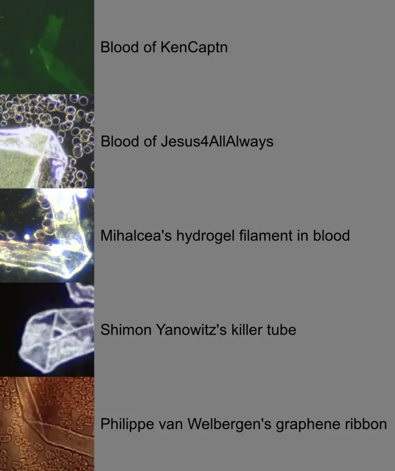
KenCaptn said that the treatment he got in Japan was very expensive,
but he had no choice because he would've died if he didn't get the
treatment: [https://

He tweeted: "This clot issue possibly could turn
into the biggest problem that humanity has ever faced. Think youre safe
because youre unvaxxed? Think again. There are violently ill people here
that are unvaxxed in Japan covered in microclots being treated."
[https://
KenCaptn also said that stem cell growth factors repair demons: [https://
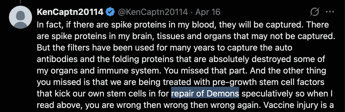
He said that blood clots are "called fibril or
amyloids": [https://

He claims that his life was earlier saved by Mary Talley Bowden who
suggested him to double his dose of ivermectin and nattokinase: [https://
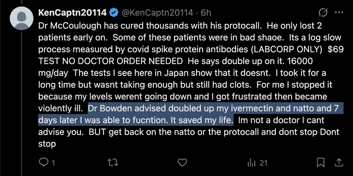

Even after he had received the DFPP treatment, he still said that he
is afraid to ever get off ivermectin: [https://
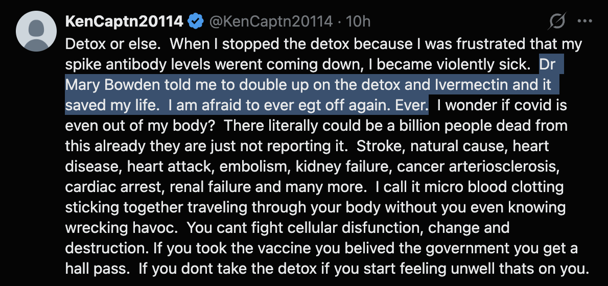
Now McCairn seems to have come up with a new explanation for why some
of his supposed clots have a flat and not cylindrical cross-section,
which is that "veins are flat and
non-cylindrical": [https://
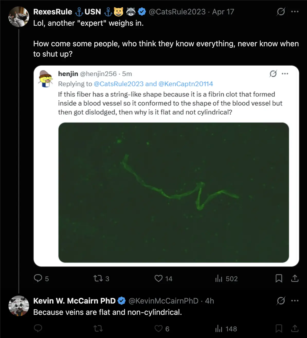
So he seems to still be saying that the shape of the clot is explained by the clot being an intravascular cast. ChatGPT said:
Short answer: generally no - a fibrin clot that detaches from a vessel does not reliably preserve the vessel's cross-sectional shape, especially not as a stable, flat "ribbon."
Fibrin networks are viscoelastic and deformable rather than rigid. Once a clot is dislodged and no longer constrained by blood flow and vessel walls, it undergoes stress relaxation, elastic recoil, and internal restructuring. As a result, it can collapse, twist, or fragment, rather than maintaining a fixed molded geometry.
Although veins can be partially collapsed or non-circular in vivo, particularly at low pressure, their lumen remains a three-dimensional, compliant structure rather than a consistently flat channel. After removal from the body, the loss of internal pressure and surrounding tissue support leads to further deformation. Additional handling and fluid forces during extraction can significantly alter the clot's shape.
There are cases where biological material approximates a lumen, such as airway casts or some post-mortem clots, but these typically appear as irregular cylindrical or branching structures. They do not present as long, uniformly flat ribbons.
A persistent flat, ribbon-like morphology is therefore more consistent with ex vivo artifact, mechanical compression, surface-interface effects, or non-fibrin material. It is not well explained by simple intravascular molding of a fibrin clot.
Overall, while a clot may transiently resemble the vessel it formed in, it does not stably retain that geometry, and a consistently flat cross-section is not what would be expected from a true intravascular fibrin clot.
When I asked RexesRule why KenCaptn's fiber was flat and not
cylindrical, she didn't answer my question, but she just said that I was
a troll, and that McCairn and his patients are heroes who don't deserve
trolls in their threads: [https://
McCairn showed me the image below from a paper by Pretorius and Kell,
where the bottom right panel appears to show a segment of a string-like
clot with a diameter of about 10 µm. But it's not clear how long the
structure is or if it has uniform width throughout its length, even
though the width of the structure does seem to taper off suddenly
towards the right side of the image. The structure doesn't look flat,
but it's not clear if other parts of the structure would look more flat.
The structure is also not uniformly fluorescent like some of McCairn's
fibers have been: [https://
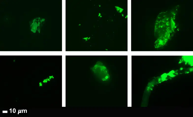
McCairn also found an Indian paper that featured a few images of
structures with a string-like shape that the authors described as
microclots: [https://
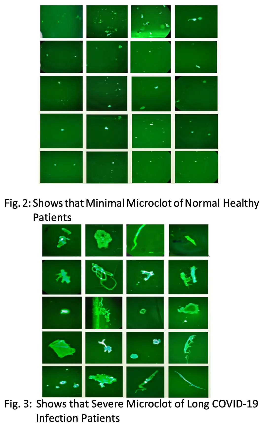
However the images in the Indian paper don't even have scale bars, so it's difficult to tell if the diameter of the string-like structures is consistent with textile fibers or not. And the elongated structures seem to have a more variable diameter than the fibers in McCairn's images, and they have a gel-like appearance, and they don't seem to be flat. The authors didn't describe analyzing the composition of the structures in order to determine if the structures were even clots made of fibrin. And the paper doesn't seem very credible, because it was written in broken English and published in a predatory journal for tropical medicine even though the paper was not about tropical medicine.
McCairn also found a paper from 2025 where the authors described a
method for quantifying the number of microclots associated with long
COVID in blood samples, which included the figure below where the top
left panel shows an elongated structure with a diameter of about 20 µm:
[https://
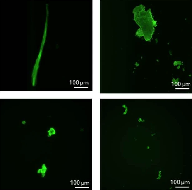
However the elongated structure above looks more cylindrical than flat, and it doesn't have any sharp corners similar to 90 degree paper folds. The structure doesn't seem have the kind of twists that are called convolutions in cotton, even though the image has such low quality that it's difficult to discern the shape of the clot. The structure also appears to have a fairly high degree of variability in diameter along its length, so that the diameter is tapered off towards the top of the image. And like in the case of the Indian study, the authors of the study did not describe determining if the structures in the images were actually made of fibrin, or doing any kind of filtering to remove non-fibrin structures from the blood.
Next McCairn found the figure below from a paper published in Nature
where the authors quantified the amount of fibrin microclots in 84
children and young adults. Like in the case of the other papers, the
authors didn't describe determining the composition of the structures to
verify if they were actually clots made of fibrin. But even if the
structure on the right is actually a clot made of fibrin, it doesn't
look flat, it has quite a bit of variability in diameter along its
length, it doesn't have sharp "paper
fold"-style corners, and it doesn't have the kind of lengthwise
fold running along the middle as the fiber McCairn found from Lyndsey's
blood: [https://
Lyndsey House showed that her plasma was more reddish before she got
McCairn's DFPP treatment and more yellowish after the treatment: [https://
When I asked what would explain the change in color, ChatGPT said:
What you're seeing actually makes physiologic sense for pre- vs post-DFPP (double filtration plasmapheresis).
A more reddish or orange plasma before DFPP typically reflects a higher concentration of circulating components that affect color and turbidity. Before DFPP, plasma contains higher levels of lipoproteins (especially LDL, VLDL, and possibly chylomicrons), immunoglobulins, and other large plasma proteins. These components can give plasma a darker, more orange or slightly reddish hue due to increased light scattering and overall density. After DFPP, many of these larger molecules are selectively removed, so the plasma appears clearer and shifts toward a more typical yellow or straw color.
Mild hemolysis can also contribute. If the pre-procedure sample has even slight red blood cell breakdown, free hemoglobin in the plasma can create a pink to reddish tint. This effect may be reduced after DFPP due to removal processes and dilutional effects. It's worth considering whether the pre sample had any handling factors that might have introduced minor hemolysis.
Plasma color is also influenced by bilirubin and concentration effects. Normal plasma is straw yellow, deeper yellow or orange can reflect higher bilirubin or increased concentration of solutes, and pink to red suggests hemolysis. After DFPP, the plasma is effectively "cleaned" and somewhat diluted, which tends to lighten its appearance.
Finally, hemoconcentration prior to the procedure can play a role. If the patient was relatively volume-depleted beforehand, plasma components would be more concentrated, contributing to a darker appearance. Fluid shifts during and after DFPP can make the post sample look lighter by comparison.
Overall, the pre-DFPP plasma appears more red or orange primarily because it contains higher levels of lipoproteins and proteins, with possible minor contributions from hemolysis or concentration effects. The post-DFPP plasma looks clearer and more yellow because those larger, color-influencing components have been reduced.
Lyndsey and another one of McCairn's patients reported reduced
numbness in their toes after getting the DFPP treatment, which might be
an outcome of improved perfusion to the toes: [https://
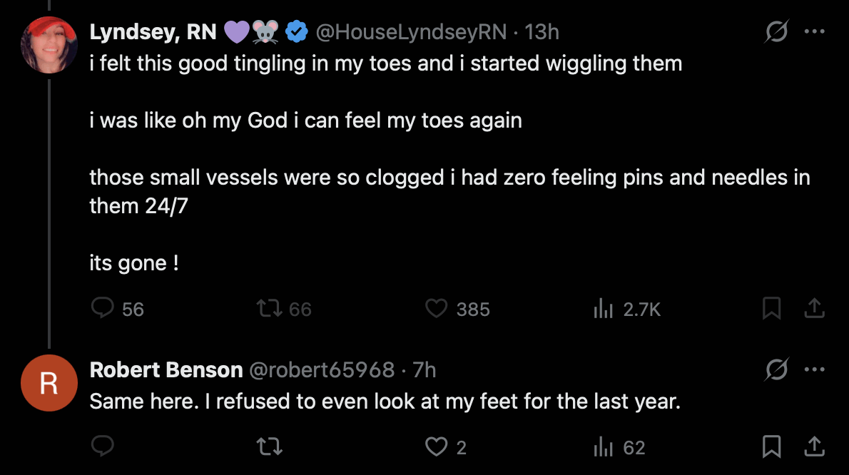
I was wondering that if DFPP doesn't actually induce microclots lodged in the capillaries to break loose, then how could it improve perfusion to the toes? Grok speculated it might be if DFPP reduces the viscocity of blood:
Fibrinogen is a key high-molecular-weight plasma protein that dramatically increases viscosity (especially in low-shear microcirculation). DFPP removes 50-88% of fibrinogen per session or cumulatively (along with other contributors like IgM, α2-macroglobulin).
Lower viscosity directly increases capillary flow rate per Poiseuille's law: flow ∝ (pressure × radius⁴) / (viscosity × length). In narrow capillaries, even small viscosity drops yield large perfusion gains. This improves oxygen exchange without needing to dissolve every lodged clot. Studies on plasmapheresis in hyperviscosity syndromes confirm this: reduced plasma viscosity enhances capillary perfusion, shear-dependent vasodilation (via endothelium-derived relaxing factors), and tissue oxygenation.
Result: Better overall microcirculatory flow, even around partial obstructions, leading to the "rejuvenating effect" described in patient reviews.
A paper about H.E.L.P. apheresis by Beata Jaeger et al. said: "In principle, the decreased fibrinogen concentration
causes rheologically significant effects and facilitates oxygen
exchange. Plasma viscosity is reduced by an average of 19%, and
erythrocyte aggregability is significantly decreased by 60% (33). In addition, the vascular endothelial growth
factor (VEGF) and nitric oxide (NO) release are favorably influenced (33). The improvements have also been demonstrated
for cerebral blood flow in the cardiac patients, where they profit from
a 63% increase in the CO2 reserve capacity (54)." [https://
The reduction in peripheral neuropathy might also be explained by a
neurological and not circulatory mechanism. Plasmapheresis is employed
as a treatment for chronic inflammatory demyelinating polyneuropathy
(CIDP), which is an autoimmune condition that
affects the myelin sheath around peripheral nerves. Plasmapheresis
filters out antibodies in plasma that attack the nerves, even though the
effect of the treatment typically lasts for only a few weeks, so the
treatment has to be repeated frequently. [https://
A paper from 2011 said: "Based on 2 Class I
studies, plasmapheresis is established as effective in the short-term
treatment of CIDP; both studies showed the beneficial effect is not
sustained, with worsening beginning 1-5 weeks after last plasmapheresis
treatment." [https://
When I asked what other explanations there might be for the reduced numbness in the toes after the DFPP treatment, Grok produced this list of 4 possible explanations:
1. Reduction in systemic inflammation and neurotoxic/inflammatory mediators (non-CIDP pathway)
DFPP removes cytokines (e.g., TNF-α), chemokines (e.g., RANTES), oxidized LDL, complement components (C3/C4), and other pro-inflammatory molecules that can directly sensitize or damage peripheral nerves or cause small-fiber neuropathy symptoms.
In patients with metabolic or non-metabolic peripheral neuropathy (including cases with environmental toxin burden), DFPP has been shown to significantly lower serum CRP, TNF-α, eosinophilic cationic protein, and fibrinogen while simultaneously removing heavy metals/pesticides - correlating with clinical sensory improvement.
Similar rapid anti-inflammatory effects are reported in autoimmune encephalitis and other neuroinflammatory states treated with DFPP, where neurological function (including sensory symptoms) improves even without classic demyelinating neuropathy.
This is a neurological (rather than purely circulatory) mechanism: the nerves themselves are less irritated or inflamed, independent of blood flow.
2. Improved rheology beyond simple viscosity (erythrocyte behavior, endothelial function, and microcirculatory dynamics)
Even without physically "breaking loose" lodged microclots, DFPP alters blood flow properties in ways that enhance capillary perfusion and tissue oxygenation:
It reduces erythrocyte aggregability/rouleaux formation (by ~60% in analogous H.E.L.P. apheresis) and lowers plasma viscosity more broadly than fibrinogen removal alone. This increases shear stress on vessel walls, triggering endothelium-derived relaxing factors (nitric oxide, VEGF) that promote vasodilation.
In conditions like arteriosclerosis obliterans (ASO) or diabetic foot syndrome - where patients classically report toe numbness, coldness, and pins-and-needles - DFPP is an established therapy precisely because it improves lower-limb microcirculation and sensory symptoms via these rheological changes.
Long-COVID apheresis studies (using similar filtration) document disappearance of fibrin fibers and rouleaux on dark-field microscopy alongside symptom relief, showing flow improvement without requiring clot dissolution.
Poiseuille's law still applies, but the effect is amplified by better red-cell deformability and reduced cellular aggregation, allowing plasma to "squeeze" through partially compromised capillaries more effectively.
3. Removal of other hyperviscosity or pro-aggregatory factors (e.g., cryoglobulins, lipids, immune complexes)
DFPP is highly effective for cryoglobulinemic vasculitis or hyperviscosity syndromes, where large proteins cause sluggish flow and neuropathy; sensory symptoms (including distal paresthesias) often resolve rapidly after sessions.
Even in non-cryoglobulinemic cases, it removes oxidized lipids and immune complexes that promote endothelial dysfunction or microvascular sludging - effects documented in long-COVID cohorts and peripheral-artery-disease patients.
Grok said that blood viscosity would typically return close to a normal level in 3-6 days, fibrinogen in 2-5 days, cytokines like TNF-α and IL-6 in about 1-2 days, and oxidized LDL in 1-7 days.
The German anti-vaccine researcher NarfGb posted these comments about
McCairn's treatment protocol: [https://
Sorry to say, but this sounds like voodoo without any given evidence! Treating symptoms temporarily is not healthy, offers no better survival chances, and is nothing more than voodoo medicine.
More open questions than anything I'd suggest for treating:
How was it proven that apheresis triggered anything more than a temporal and/or placebo effect? (How was a sham-controlled effect size established to differentiate therapeutic apheresis from a temporal/ placebo response?)
How was the clearance of modRNA determined following apheresis? (Lack of pharmacokinetic data regarding the removal of synthetic transcripts from systemic circulation.)
In the event of persistence, how were integrative events and/or RNA turnover, driven by cellular homeostatic effects, excluded? (Addressing the risk of genomic integration or compensatory mechanisms masking the treatment's actual impact.)
How was the clot formation mechanistically attributed to modRNA/Spike persistence without relying on third-rate in vitro experiments that lack any reliable diagnostic or predictive value?
How was the individually dependent dosage determined, particularly concerning the administration of growth factors? (Considering the non-linear dose-response curve and the risk of biological recklessness.)
Which specific exosome formulation was utilized? (Demanding CMC, Chemistry, Manufacturing, and Controls-data: source, purity, and surface markers.)
Which specific growth factors were used, and what specific cell-line was their recombinant or biological origin? In other terms: what is "SHED"?: It is not clear if it were Mesenchymal stem/stromal cells (Dental Pulp MSCs / SHED-MSCs), Perivascular stem cells / pericyte progenitors, Neural crest-derived stem/progenitor cells, Neural stem or neural progenitor cells, Odontoblastic progenitor cells (dentin-forming precursors)
How was the individual dose-dependency established to avoid over-stimulation of signaling pathways?
By what methodology were the exosomes tracked in vivo to confirm biodistribution and target-tissue engagement?
How were off-target effects and non-specific cellular interactions rigorously excluded?
What is the clearly defined molecular mechanism of action (MoA), including validated in vivo dose-response relationships, that substantiates causality beyond descriptive reports and in vitro experiments employing recombinant spike proteins lacking physiologically relevant dosing?
Where is the supporting multi-omics data (transcriptomics, proteomics, metabolomics, lipidomics (due to using exosomes)) to provide a quantifiable biological basis?
How were all other potential etiologies, other than the "Spike protein", differentially excluded?
What is the exact N-number and stratification of the cohort regarding previous modRNA-LNP injections?
How was electrostatic membrane interference and receptor shifting excluded, which may lead to disturbed intra-cellular communication, as primary drivers for thrombotic events?
How is the absence of time-shifted feedback loops or paradoxical immune responses excluded after application that could lead to long-term exacerbation?
Without addressing dose-dependent toxicity and providing longitudinal omics-data, and a clear tier of evidence this protocol remains a speculative 'Black Box' that fails the most basic requirements of the Helsinki Declaration.
In the comment section NarfGb wrote: "There is not even any animal experiments done. But using it for people with real issues and getting paid for doing human experiments you think is a good thing?! Especially with a therapy that does not really differ from LNPs despite a little bit more biocompatible lipid composition?"
He also wrote: "Sadly: It is not even a
scientific argumentation: 'BUT they look and feel
great.' I feel also great smoking crack or taking
steroids." And he expressed concern how McCairn did not provide
numeric data to quantify the effect of his treatment, but he presented
evidence like an infomercial-style image where the patient looked
happier in the "after" picture than the "before" picture. [https://
NarfGb also wrote: "And in the first line I am more pissed about the fact that he sells this protocol as a mirrage cure than to say it's pure experimental and we don't know by now what we're doing. This is the part really freaks me out." I agree with his concern, and furthermore McCairn has been fearmongering about the clots to the extent that some of his followers now believe that the clots are leading to human extinction. One of his patients said that the clots might be the most important issue humanity is facing, and that everyone is dying from the clots, and that he would have died if he didn't get McCairn's treatment. The patient also speculated that the stem cell growth factors "repair demons", which makes it seem like he didn't understand the purpose of the treatment well enough to have informed consent.
NarfGb next published a second post about the McCairn's protocol.
[https://
I'm not saying that abnormal, fibrinolysis-resistant clots don't exist. The fluorescence signal is reproducible. But the term "amyloid" is overinterpreted and unproven.
I replied:
I agree, and the "amyloid" terminology fuels McCairn's fearmongering about how the vaccines are causing a prion zombie apocalypse.
Two years ago Baxter Dmitry from The People's Voice published an article that said: "According to Dr. McCairn, contagious prions released by Covid jabs behind the explosion of disease around the world and explain the mechanism for phenomena including 'silent hypoxia', the formation of white fibrous clots, the sudden deaths of athletes, turbo CJD, turbo cancer, amyloidosis, and the explosion of neurological diseases." [https://
thepeoplesvoice. ]tv/ neuroscientist- contagious- prions- from- covid- jabs- are- transfecting- the- unvaccinated/
Then NarfGb told me:
You knew, that you'll find in almost every protein amyloidogenic peptide sequences?
Tzotzos S, Doig AJ. Amyloidogenic sequences in native protein structures. Protein Sci. 2010 Feb;19(2):327-48. doi: 10.1002/pro.314. PMID: 20027621; PMCID: PMC2865711.
That was the best joke for the panic narrative: using the worst sequences to sell a panic narrative and distract from the injections.
I told him that was a great point, and I hadn't explored that angle before. Grok said:
Amyloidogenic or aggregation-prone sequences (APRs) are very common across proteins. These short segments, typically 5-15 residues long and often hydrophobic with high β-sheet propensity, occur frequently in polypeptide chains. Predictive tools such as TANGO, WALTZ, and AGGRESCAN, along with large-scale proteome analyses, indicate that APRs appear on average at least once every ~50-100 amino acids in typical protein sequences. In globular proteins, roughly 15-20% of residues fall within such predicted aggregation-prone regions, though these are usually buried in the hydrophobic core or stabilized by native folding to prevent uncontrolled aggregation. [https://
www. ]nature. com/ articles/ s41467- 020- 17207- 3 Proteome-wide surveys confirm this high prevalence. In a comprehensive analysis of plant proteomes spanning 75 species and more than 2.9 million protein sequences using the WALTZ algorithm, more than half of all proteins in each proteome contained at least one predicted amyloidogenic region, with a median of around 25% of proteins after applying stricter filters. [https://
www. ] Similar patterns hold in other organisms, including bacteria and eukaryotes. Many proteins - perhaps most - harbor the potential to form amyloid-like structures under permissive conditions such as fragmentation, denaturation, high local concentration, or specific environmental stresses.mdpi. com/ 1422- 0067/ 18/ 10/ 2155
A user that promotes Miles Guo said that the CCP is using a protocol
similar to the McCairn protocol but with fresh blood taken from PLA
military boys: [https://
The account has a similar profile picture and banner image as two
other accounts that promote Miles Guo: [https://


A paper about bleeding risk associated with DFPP said: "Notably, although most coagulation factors typically
normalize within 48-72 h after DFPP treatment, fibrinogen has a long
biological half-life of about 3.5 days and incomplete recovery at 48 h
post treatment is the norm [16]."
[https://
Jordan Vaughn in Alabama offers a similar services of testing for
microclots as McCairn. One of his patients posted this tweet that showed
a report they received from Vaughn, which said: "Micro-clots come in all shapes and sizes. You may also
see long, string-like appearing objects in your pictures. These are
Endothelial cast and are associated with endothelial damage and
inflammation. This is a normal finding for long-COVID patients."
The report included three images of elongated clots, even though none of
them looked flat, and all had a variable diameter: [https://
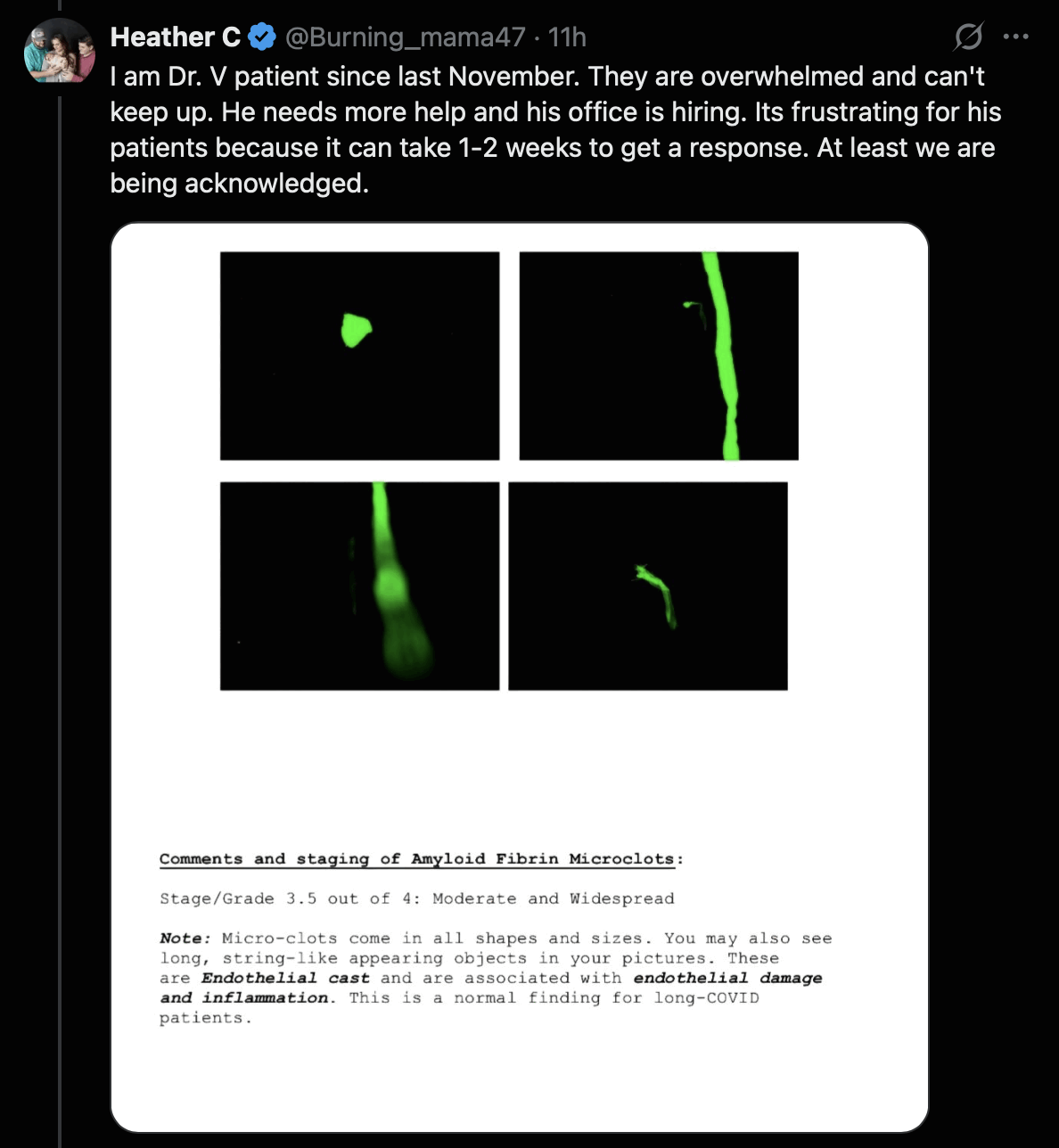
Apheresis Center in Cyprus also offers testing for microclots
associated with long COVID. The website of the center includes these
images of microclots, which all have a globular and not elongated shape:
[https://
Peter McCullough scams people by selling a test for spike antibodies,
and then he tells people with a high level of antibodies to buy his
products in order to "detox" the spike
protein, as if a high antibody level would be a sign that people have
spike protein left over in their body. However in reality the presence
of antibodies for the spike protein is generally a sign that immunity to
COVID has been triggered by a vaccine or a natural infection. McCairn
replied to a tweet about McCullough's test by saying: "Our patients show significant drops once treatment
starts, and extends out for all tested patients. It's an indirect
measure of the underlying pathology though." [https://
But how is it a symptom of a "pathology" if someone has antibodies for the spike protein? If McCairn's plasmapheresis treatment actually resulted in the permanent removal of spike antibodies, it might make his patients more susceptible to COVID, which would seem like a downside of the treatment. But of course the immune system will replenish the antibodies soon after the treatment. Antibodies are produced by memory B cells, which mainly reside in the lymph nodes, spleen, tonsils, and bone marrow. And even though a small part of memory B cells are circulating in blood, memory B cells that are removed from the blood still get replenished through a mechanism where the cells make copies of themselves through cell division.
One of McCairn's patients said that he was taking McCullough's Spike
Detox supplements in order to reduce his level of spike protein
antibodies, as if the antibodies would've somehow been harmful. [https://
One of McCairn's patients posted the images below as evidence that
her treatment was successful, because her blood looked more "stringy" before the treatment but more "smooth" after the treatment: [https://
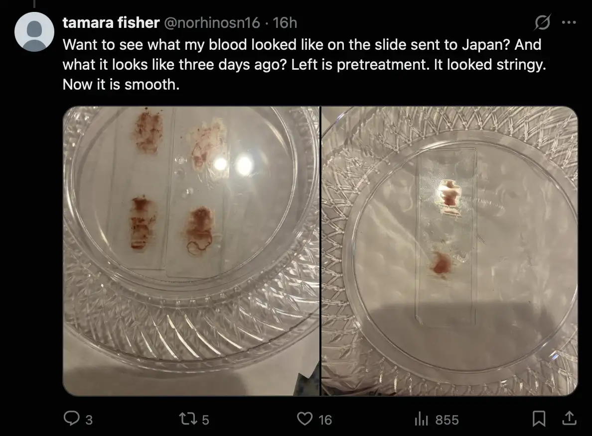
Her images were turned into an infomercial-style image by the memetic
warrior from naval intelligence: [https://

In June 2025 McCairn did an experiment where he mixed fibrinogen with
ThT, heated it up to 42 degrees, and shook the mixture, and as a result
he claimed he was able to create the fluorescent fibers below. He said
that "using heat and shaking, we've managed to make
the fibrinogen aggregate into an amyloidogenic form". [1:39:39] And he said: "In this
instance I've been able to make fibrillar forms twisted reactive profile
that looks very similar to what we're seeing in the blood. And again,
what's the critical factor here? Is it the temperature? Is the
Thioflavin intrinsically causing a reaction to the fibrinogen? Is the
concentration of fibrinogen too high?" [1:29:43] He was surprised to find fiber-like
structures that looked similar to the so-called "amyloid fibrin aggregates" he found in blood
samples, even though his experiment was supposed to be the control run
where he just looked at regular fibrinogen and he didn't apply any
special procedure to form amyloid fibrin, like mixing in LPS or spike
protein peptides with the fibrin. But he didn't rule out the possibility
that the fibers were not made of fibrin, but they were just contaminants
on his slide: [https://
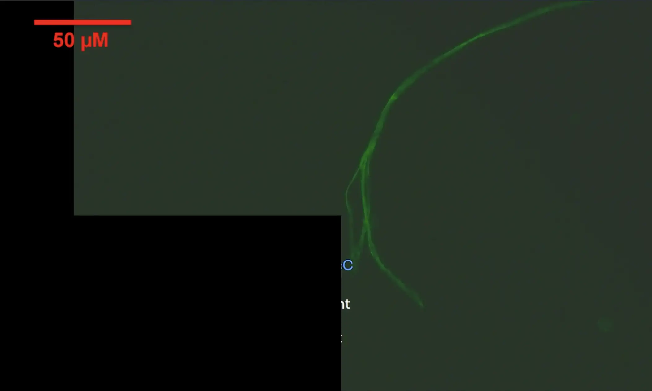
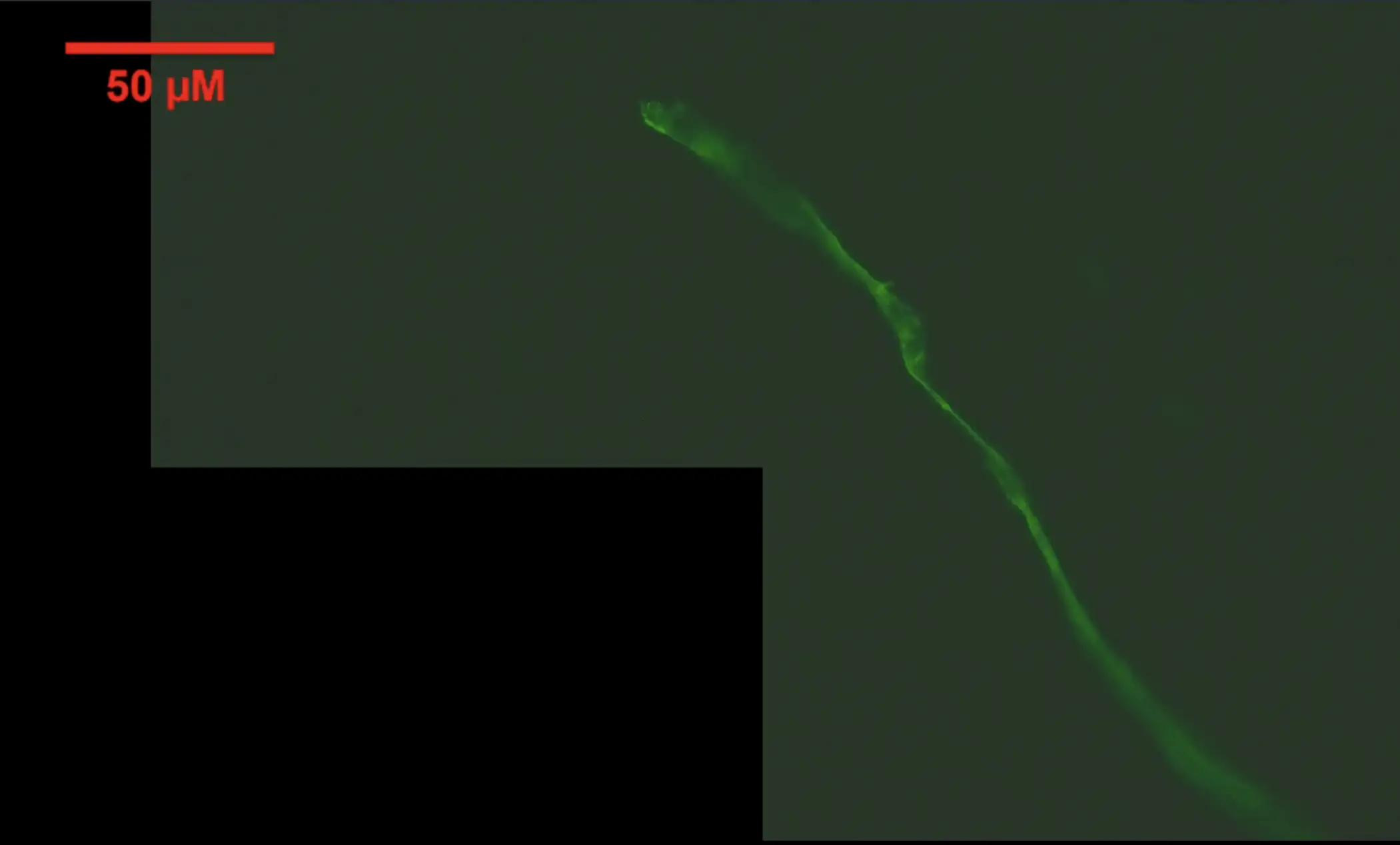
At time 1:18:12 he said "What I've done to try to remove confounders that - fibers, dust, et cetera that could get to our preparation - so every sample that we're using here, has been passed and filtered through a - basically anything larger than a 100 micrometers would not pass the filter". But he didn't mention that fibers from dust narrower than 100 µm might still pass through the filter.
He should've also repeated the experiment without the Thioflavin, so he could've seen if he found similar fibers that were autofluorescent without Thioflavin.
When I asked McCairn why the fiber in KenCaptn's blood had a flat shape, McCairn said it was because blood vessels are flat. But the fiber in the second image above also has a flat shape, even though the fiber didn't form inside a blood vessel.
In 2025 when Ian Musgrave said that the fiber in the 3-year-old's
blood was cellulose and McCairn got a false positive with ThT, McCairn
said that in order to eliminate false positives, "I
run antibodies, use SEM/EDX, and Raman on samples". [https://
In 2012 the CDC published a study where they analyzed supposed
Morgellons fibers but found them to be consistent with textile fibers or
cellulose. The IR spectroscopy result were also consistent with
cellulose, but I have never seen McCairn publish similar spectroscopy
results of his supposed fibrin clots: [https://
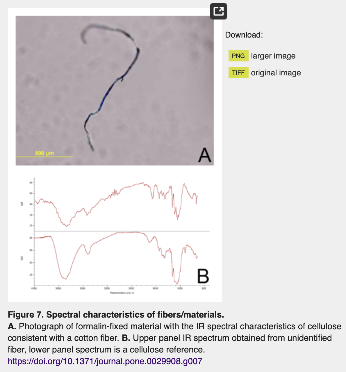
A Twitter user whose display name is "china
Victims of Sinovac COVID-19 Vaccine" was inspired by McCairn to
get DFPP treatment in China. But he said "DFPP is
completely ineffective; everything rebounded completely after four days.
What a tragedy": [https://
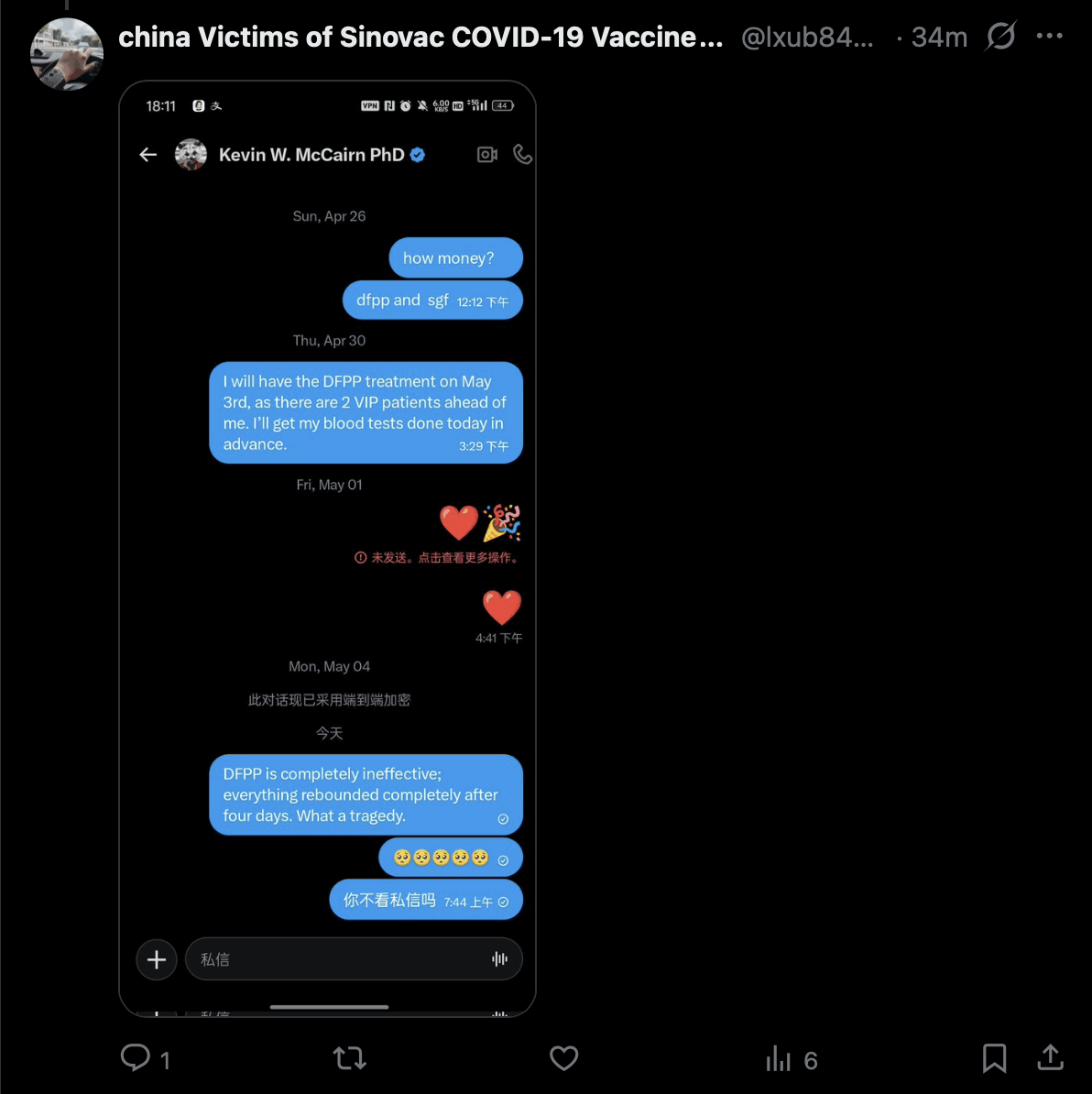
He also said "DFPP has left my body weakened, and
it delivered zero therapeutic effect". He was confused about
whether he should've gotten DFPA or DFPP, but it's likely the results of
DFPA wouldn't have been much different: [https://
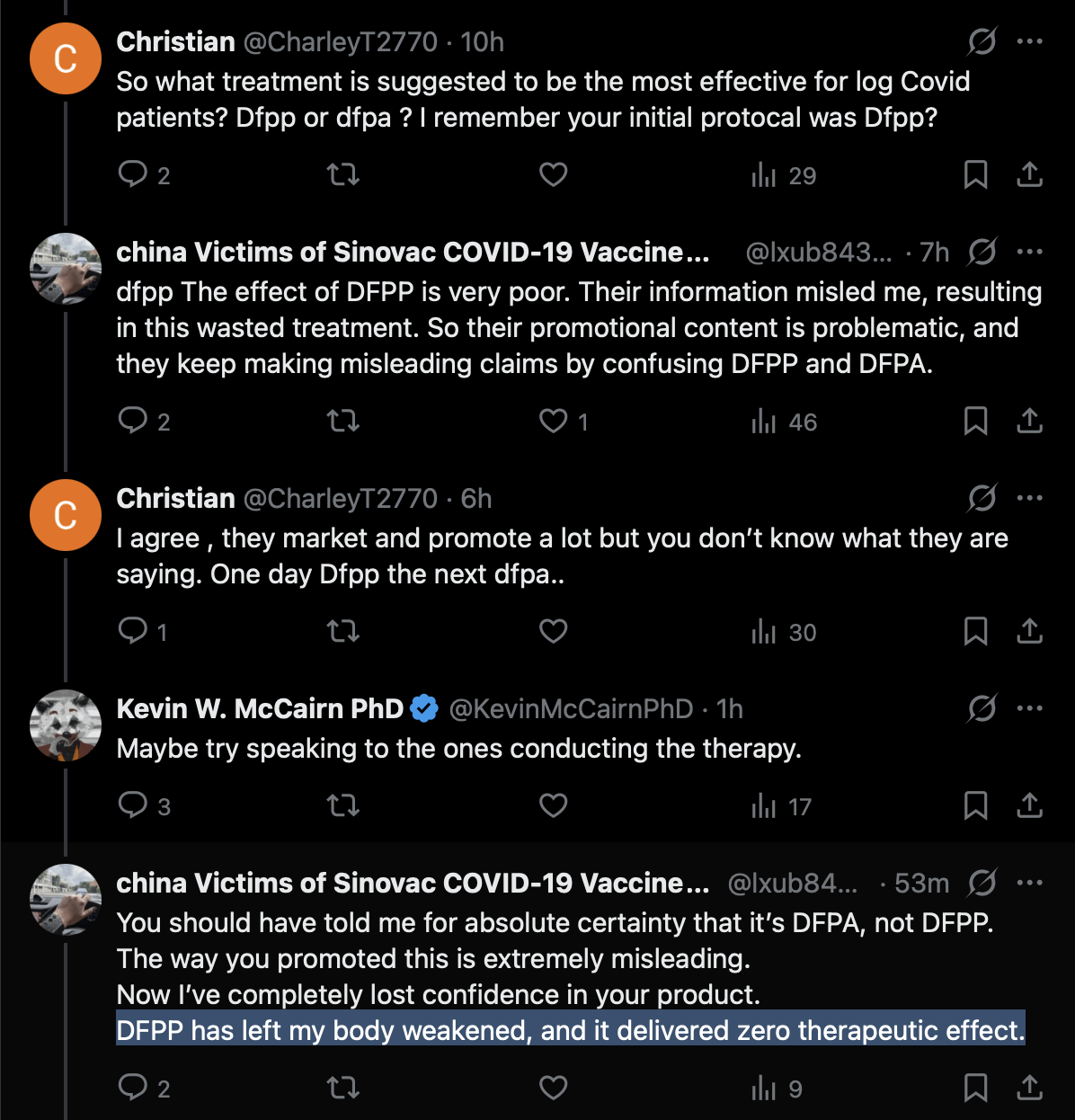
McCairn seems to have blamed the lack of success on getting DFPP
instead of DFPA and stem cell growth factors, or on using unclean donor
plasma. The Chinese guy was told by a Japanese supporter of McCairn that
DFPP should be fine: [https://
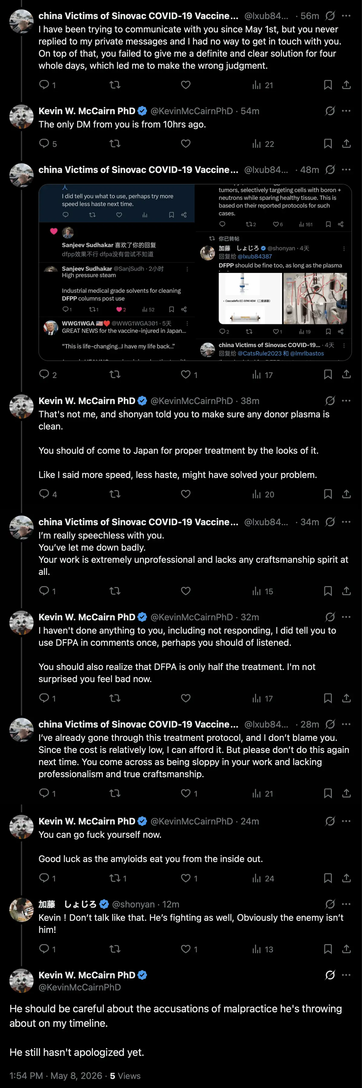
Despite what McCairn said in the last tweet above, the Chinese user
didn't accuse McCairn of malpractice, but only of not replying to his DM
and not making clear enough if his protocol used DFPP or DFPA, and
McCairn later admitted he wrong that the Chinese user hadn't sent a DM
to him. [https://
Another Chinese user responded that RexesRule and CoyoteSanctuary
also spoke about DFPP and not DFPA, and he thought they were part of
McCairn's marketing team: [https://
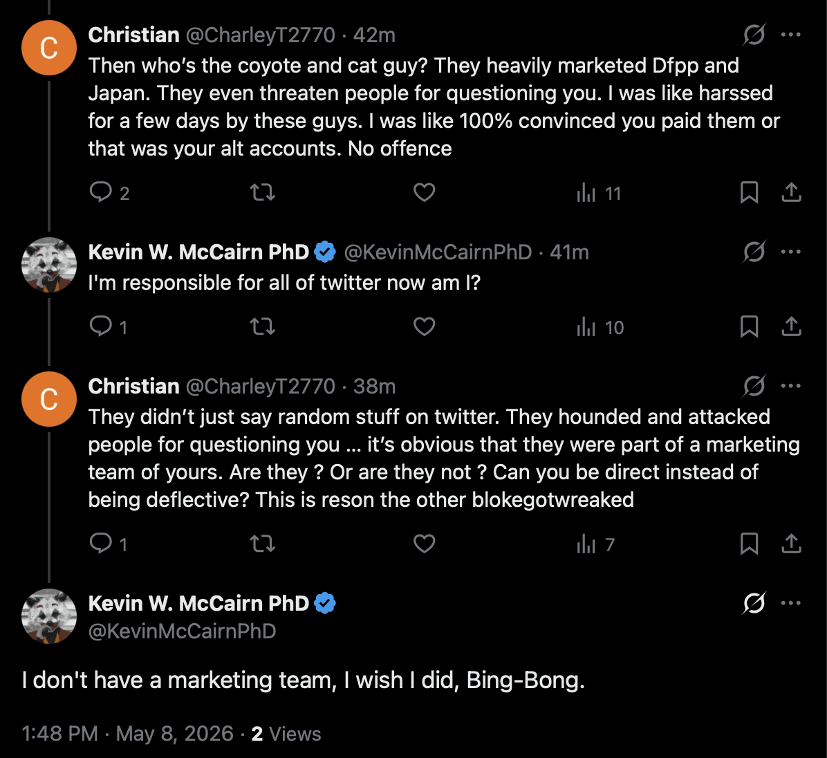
McCairn's patient mycityapartment wrote about the treatment that
"I have experienced significant improvement that
anticoagulants did not/could not provide". [https://
When I asked two of McCairn's patients if they had a rebound in
symptoms similar to the Chinese guy, one of the patients said: "No I'm slowly improving across all symptoms including and
especially neuropathy. 6 weeks from when I got home." [https://
But anyway, it was understandable for the Sinovac victims guy to mix
up DFPP and DFPA, because the website of the hospital that provides the
treatment says: "Edogawa Hospital's program is
presented around Dual Filtration Plasmapheresis, also described as DFPP
or DFPA in patient materials". [https://
A Japanese guy who did McCairn's blood test said this elongated
structure was in his blood. The structure is fairly short, it has a
variable diameter, it doesn't look too flat, and it doesn't have sharp
folds, so it might actually be made of fibrin: [https://
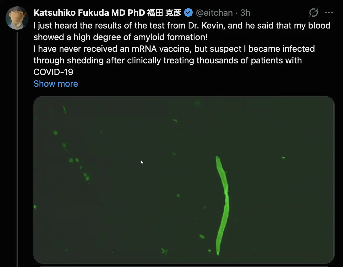
But McCairn still doesn't seem to be doing any procedure to determine the composition of the clots, like spectroscopy or immunostaining for fibrin antibodies.
One of McCairn's patients from May 2026 said he didn't notice any
improvement after the DFPA treatment: [https://
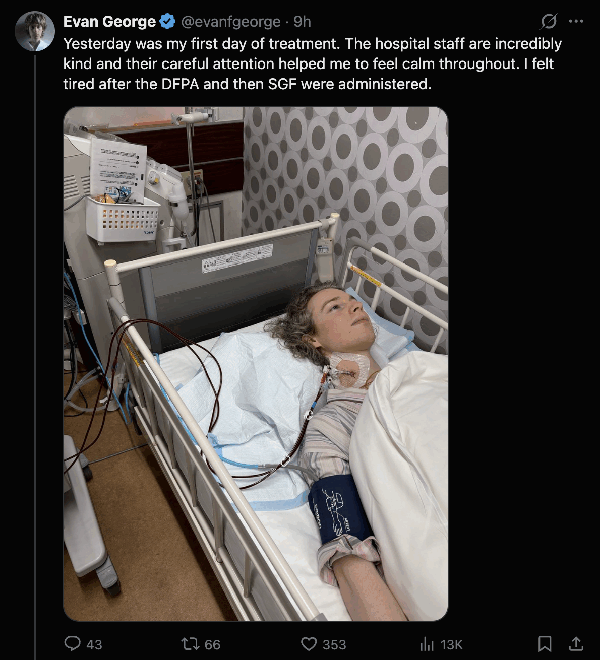

A few days later when he was asked about the effect of the SGF
treatment, he said: "It's subtle for me, but I
definitely have been just a little bit more functional. I'm able to do a
bit more before feeling sore but I still will start to feel symptoms if
I push." [https://
Resia Pretorius quoted my tweet and wrote: "We
and others looking for fibrinaloid microclot complexes, always do
analysis on fresh blood plasma samples or plasma spun and stored at -80.
We do not support dried sample analysis and any shipment at room temp.
One must also be careful, as tissue paper fibers can show
autofluorescence. However, we cannot comment on this exact
photo." [https://
Grok said: "Tissue paper fibers fluoresce under UV light primarily due to optical brighteners (also called fluorescent whitening agents or FWAs) added during manufacturing. These colorless compounds, often stilbene derivatives or distyrylbiphenyl types, absorb invisible ultraviolet radiation (typically 340-370 nm) and re-emit it as visible blue light (around 400-450 nm) through fluorescence." Similar brighteners are also used in white textiles.
Tissue paper fibers look somewhat similar to cotton fibers and other
cellulose fibers: [https://
This shows toilet paper fibers: [https://
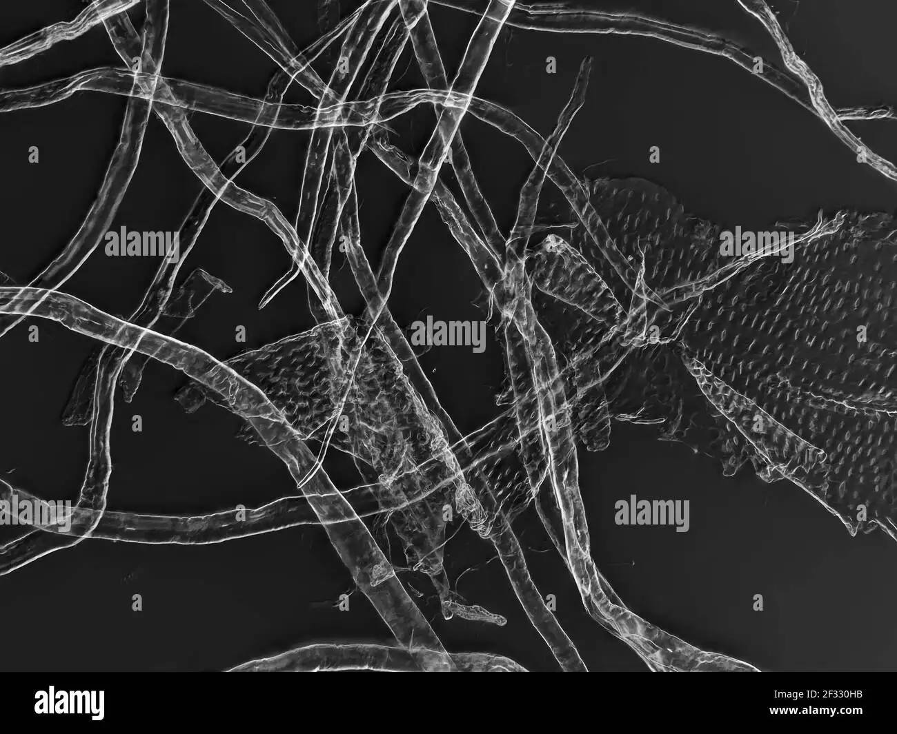
Resia Pretorius also posted these answers to replies to her tweet:
In June 2026 Mary Talley Bowden did an interview with McCairn and his
patient Ken Evans, whose username on Twitter is KenCaptn20114. [https://
At time 13:33 Ken Evans said: "We already know that we have spike proteins, we already know we have amyloid. We're just trying to survive. And that's the part of this whole Japan thing. I was not prepared for seeing how ill these people were. If you have a heart and you see these people, it will make you so mad that somebody is in this condition. And we all know that we had amyloids. They were stuck in all our tissues. And in my body, they were opening up in the sores. I was sneezing blood, I was coughing blood, blood in my feces, my stomach, my esophagus, liver, lungs, kidney, fibrosis, my swollen spleen, my heart. Every part of my body was absolutely devastated."
But how did he know he had "amyloids" in other tissues besides his blood? As far as I know, McCairn only tests for the presence of the amyloid fibrin clots in blood, and it wouldn't make sense for the fibrin clots to even exist in other tissues.
Ken Evans also seems to think that because he still has antibodies to the spike protein, it means that his body is full of spike protein coming from the vaccines. At time 16:58 he said that after he got McCairn's treatment, "My spike proteins went down 25%. So the treatment for me not only saved my life, but it pulled a lot of autoantibodies, out misfolding proteins, spike proteins, and all of those blood clots." So he seemed to think the reason why his level of spike protein antibodies was reduced by 25% was because he still had spike protein from the vaccines circulating in his blood which was removed by McCairn's treatment, which doesn't make any sense.
At time 47:49 Bowden asked: "And just to be clear, normally in a normal blood you would not see anything lighting up at all?" Then McCairn showed the image below and said: "I mean you can see something. So look, if you look at that image that's on the screen, you can see smaller - like tiny microdots - that cluster around the image. Every sample will have that - because as the protein dries and desiccates on the slide, and there are some proteins and molecular species that are just fluorescent in and of themselves - but the size differential is what makes them different, okay. The fibrin causes large amyloidogenics twisting structures." Earlier McCairn has given the impression that the smaller fluorescent blobs are also be made of "amyloid fibrin", but now he seemed to suggest that the small blobs are not necessarily made of fibrin, but a characteristic of fibrin aggregates would be a large size or a twisting shape.

At time 1:26:04 Ken said: "People that are taking
the vaccine is safe trail are ignoring Pfizer's own statistics. They
admitted their vaccine didn't work. They admitted that it killed 82% of
the pregnant women in studies." He misunderstood a set of
preliminary data from the CDC's V-safe study, where at a time when
almost no participants had yet completed a full-term pregnancy and only
about 4% of pregnancies had completed, 104 out of 127 completed
pregnancies had ended in a miscarriage (and not in
the death of the pregnant woman). Later after more pregnancies
had completed, the ratio of miscarriages divided by completed
pregnancies dropped to about 13%. [https://
Even Jane Ruby seemed to think that RexesRule was being paid by
McCairn to promote his treatment: [https://
The DFPA treatment is provided by Shoujirou Katou (加藤正二郎), who is the director of Edogawa
Hospital in Tokyo. He also organized a conference at the hospital where
the speakers consisted of Japanese anti-vaxxers and alternative medical
practicioners: [https://

Katou's pinned tweet says in Japanese that the COVID vaccine is "extremely harmful and useless": [https://
The reason why Katou became the director of the hospital is not necessarily because of medical competency, but because the hospital was founded by his great-great-grandfather. In Japan it's common for the directorship of a hospital to be passed down from father to son. The previous director of the hospital was Katou's older brother.
One of McCairn's patients wrote: "Japan has the
highest per capita mRNA vaccination rate in the world! According to
Dr. Kato, 3.M Japanese died in the year following the initial
rollout." [https://
The estimate of 3.89 million deaths was calculated based on a set of
FOI data which included about 4% of the Japanese population, and which
had about 150,000 deaths within a year from any vaccine dose, so the
number of deaths was then multiplied by 25.8 to estimate the number of
deaths in the whole Japan. [https://
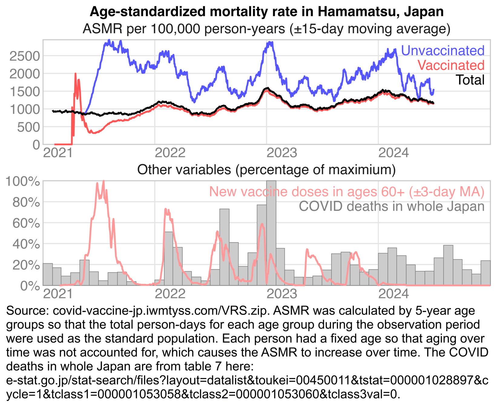
In the years 2021-2024, there were likely about 6 million Japanese people who died within a year from eating rice. But that doesn't mean that 6 million people were killed by rice. In the same way the FOI data doesn't show that 3.89 million people were killed by vaccines, but it rather shows that vaccinated people had lower ASMR than the overall Japanese population.
Now there's another cat lady who has started to say that McCairn is a
hero who deserves a Nobel Prize. She made an AI-generated video where
McCairn performs what looks like psychic surgery to "cast out amyloids": [https://
Someone who is going to Japan to get McCairn's treatment posted this
photo of a fiber in his blood. If McCairn's scale bar is correct, then
the fiber has a diameter of about 10 µm. The fiber has a fairly uniform
diameter which doesn't seem like a characteristic of a fibrin clot, but
the fiber has the kind of flat cupped body that is characteristic of
cotton: [https://
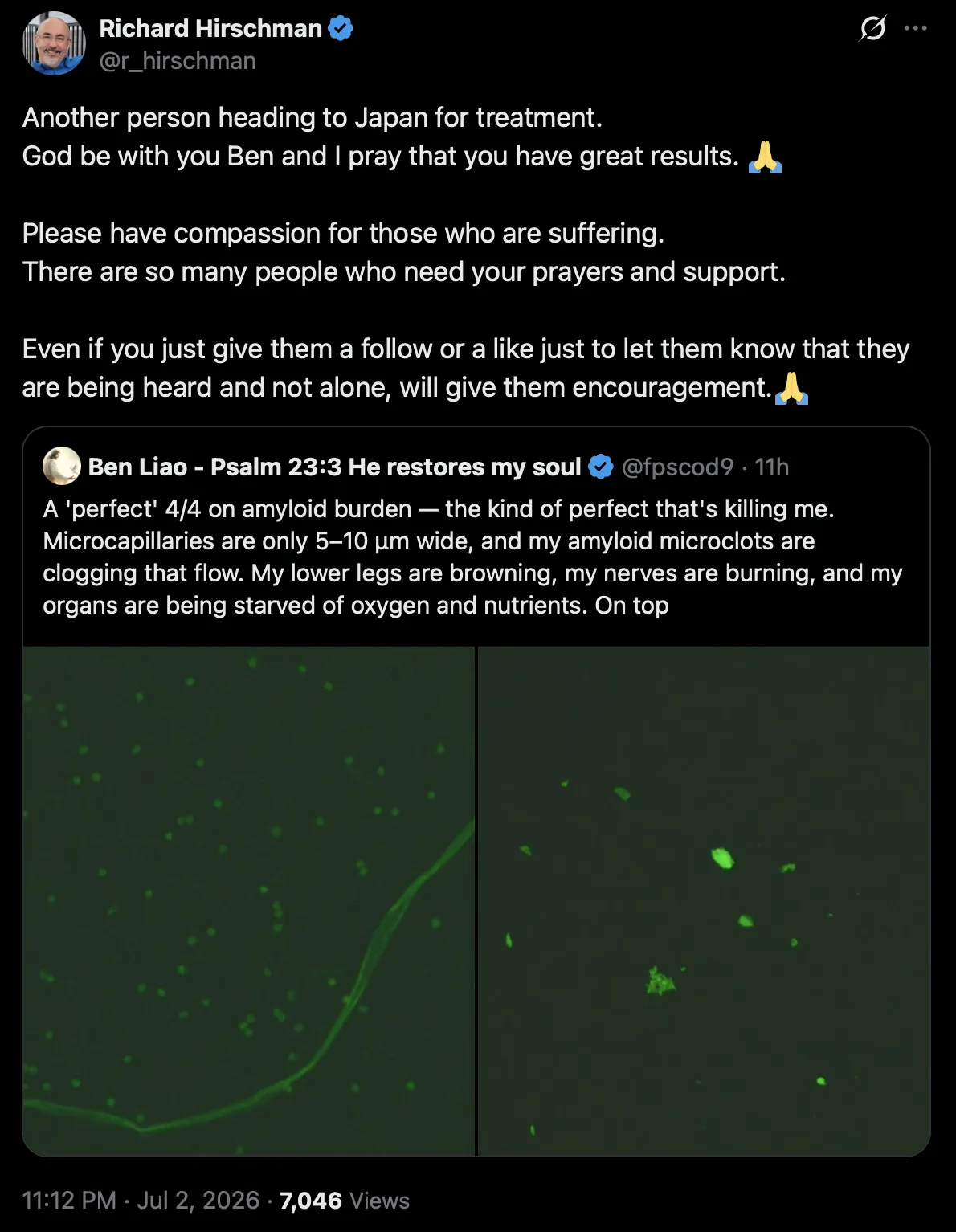
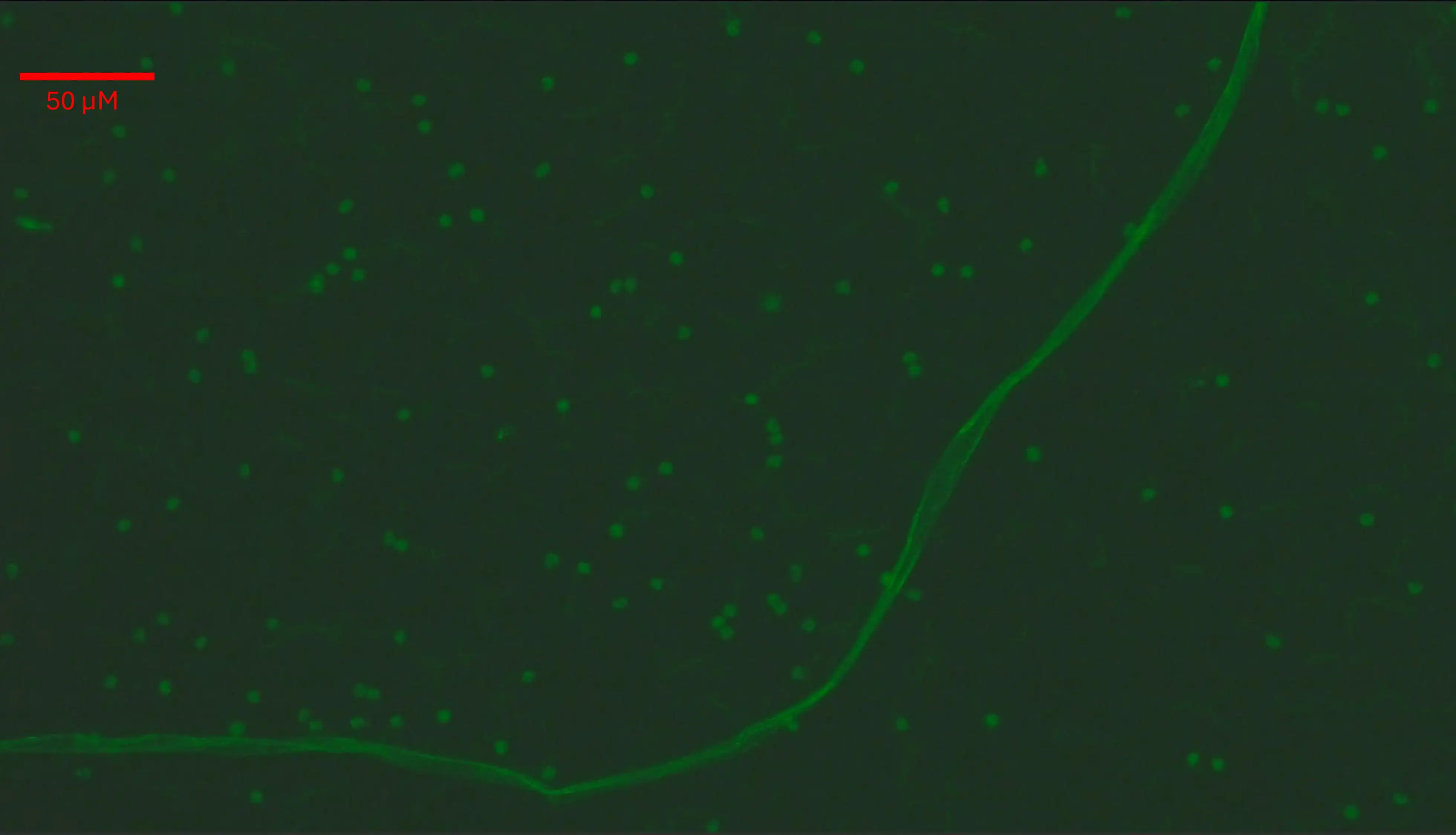
McCairn now claims that he is finding hydrogel in blood samples:
[https://
In 2023 Ana Mihalcea was similarly saying that she found hydrogel in
vaccinated blood that was developing into calamari clots: [https://
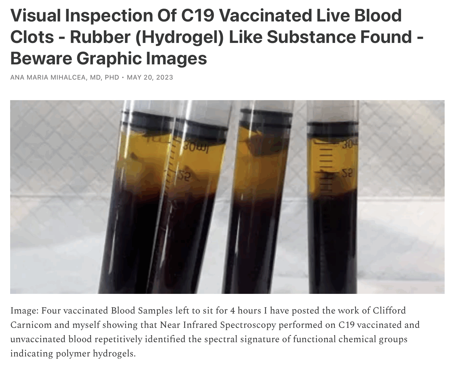